logo
AAT Bioquest

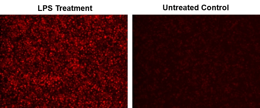
Cell Meter™ Fluorimetric Intracellular Nitric Oxide (NO) Activity Assay Kit *NIR Fluorescence Optimized for Microplate Reader*

Nitric oxide (NO) is an important biological regulator involved in numbers of physiological and pathological processes. Altered NO production is implicated in various immunological, cardiovascular, neurodegenerative and inflammatory diseases. As a free radical, NO is rapidly oxidized and there is relatively low concentrations of NO existing in vivo. It has been challenging to detect and understand the role of NO in biological systems. Cell Meter™ Fluorimetric Intracellular Nitric Oxide Assay Kits provide sensitive tools to monitor intracellular NO level in live cells. Nitrixyte™ probes are developed and used in our kits as an excellent replacement for DAF-2 for the detection and imaging of free NO in cells. Compared to the commonly used DAF-2 probe, Nitrixyte™ probes have better photostability and enhanced cell permeability. This particular kit uses Nitrixyte™ NIR that can react with NO to generate strong near-infrared (NIR) fluorescence signal. Nitrixyte™ NIR can be readily loaded into live cells, and its fluorescence signal can be conveniently monitored using the filter set of Cy5® or APC. This kit is optimized for fluorescence imaging and microplate reader applications.

Example protocol

AT A GLANCE

Protocol summary

  1. Prepare cells in growth medium
  2. Incubate cells with test compounds and Nitrixyte™ NIR working solution
  3. Add Assay Buffer II
  4. Monitor fluorescence intensity at Ex/Em = 650/680 nm

Important notes
Thaw all the kit component at room temperature before use.

PREPARATION OF WORKING SOLUTION

Add 20 µL of Nitrixyte™ NIR stock solution (Component A) into 10 mL of Assay Buffer I (Component B) and mix well. The working solution is stable for at least 2 hours at room temperature. Note: 20 µL of Nitrixyte™ NIR stock solution is enough for one plate. Keep from light.

For guidelines on cell sample preparation, please visit
https://www.aatbio.com/resources/guides/cell-sample-preparation.html

SAMPLE EXPERIMENTAL PROTOCOL

  1. To stimulate endogenous NO, treat cells with 10 µL of 10X test compounds (96-well plate) or 5 µL of 5X test compounds (384-well plate) in cell culture medium or your desired buffer (such as PBS or HHBS). For control wells (untreated cells), add the corresponding amount of medium or compound buffer. Note: It is not necessary to wash cells before adding compound. However, if tested compounds are serum sensitive, growth medium and serum factors can be aspirated away before adding compounds. Add 90 µL/well (96-well plate) and 20 µL/well (384-well plate) of 1X Hank’s salt solution and 20 mM Hepes buffer (HHBS) or the buffer of your choice after aspiration. Alternatively, cells can be grown in serum-free media.

  2. Add 100 µL/well (96-well plate) or 25 µL/well (384-well plate) of Nitrixyte™ NIR working solution in the cell plate. Co-incubate cells with test compound and Nitrixyte™ NIR working solution at 37°C for desired period of time, protected from light. Note: DO NOT remove the test compounds. Note: For a NONOate positive control treatment: Cells were incubated with Nitrixyte™ NIR working solution at 37°C for 30 minutes. The working solution was removed and cells were further incubated with 1 mM DEA/NONOate at 37°C for 30 minutes to generate nitric oxide. See Figure 1 for details. We have used Raw 264.7 cells incubated with 0.5X Nitrixyte™ NIR, 20 µg/mL of lipopolysaccharide (LPS) and 1 mM L-Arginine (L-Arg) in cell culture medium at 37°C for 16 hours. See Figure 2 for details.

  3. Remove solution in each well. Add Assay Buffer II (Component C), 100 µL/well for a 96-well plate or 25 µL/well for a 384-well plate. Note: DO NOT wash cells before adding Assay Buffer II.

  4. Monitor the fluorescence increase using microplate reader at Ex/Em = 650/680 nm (cut off = 665 nm) with bottom read mode, or take images using fluorescence microscope with a Cy5® filter.

Citations

View all 3 citations: Citation Explorer
New insights into posttranslational modifications of proteins during bull sperm capacitation
Authors: Mostek-Majewska, Agnieszka and Majewska, Anna and Janta, Anna and Ciereszko, Andrzej
Journal: Cell Communication and Signaling (2023): 1--23
Fluorescent real-time quantitative measurements of intracellular peroxynitrite generation and inhibition
Authors: Luo, Zhen and Zhao, Qin and Liu, Jixiang and Liao, Jinfang and Peng, Ruogu and Xi, Yunting and Diwu, Zhenjun
Journal: Analytical biochemistry (2017): 44--48
Inducible Nitric Oxide Synthase (iNOS) Is a Novel Negative Regulator of Hematopoietic Stem/Progenitor Cell Trafficking
Authors: Adamiak, Mateusz and Abdelbaset-Ismail, Ahmed and Moore, Joseph B and Zhao, J and Abdel-Latif, Ahmed and Wysoczynski, Marcin and Ratajczak, Mariusz Z
Journal: Stem Cell Reviews and Reports (2016): 1--12

References

View all 139 references: Citation Explorer
Pitfalls and limitations in using 4,5-diaminofluorescein for evaluating the influence of polyphenols on nitric oxide release from endothelial cells
Authors: Uhlenhut K, Hogger P.
Journal: Free Radic Biol Med (2012): 2266
Effects of moderate electrical stimulation on reactive species production by primary rat skeletal muscle cells: cross talk between superoxide and nitric oxide production
Authors: Lambertucci RH, Silveira Ldos R, Hirabara SM, Curi R, Sweeney G, Pithon-Curi TC.
Journal: J Cell Physiol (2012): 2511
Improved measurements of intracellular nitric oxide in intact microvessels using 4,5-diaminofluorescein diacetate
Authors: Zhou X, He P.
Journal: Am J Physiol Heart Circ Physiol (2011): H108
Aging negatively affects estrogens-mediated effects on nitric oxide bioavailability by shifting ERalpha/ERbeta balance in female mice
Authors: Novensa L, Novella S, Medina P, Segarra G, Castillo N, Heras M, Hermenegildo C, Dantas AP.
Journal: PLoS One (2011): e25335
Temporal and spatial correlation of platelet-activating factor-induced increases in endothelial [Ca(2)(+)]i, nitric oxide, and gap formation in intact venules
Authors: Zhou X, He P.
Journal: Am J Physiol Heart Circ Physiol (2011): H1788
Page updated on September 19, 2024

Ordering information

Price
Unit size
Catalog Number16359
Quantity
Add to cart

Additional ordering information

Telephone1-800-990-8053
Fax1-800-609-2943
Emailsales@aatbio.com
InternationalSee distributors
Bulk requestInquire
Custom sizeInquire
Technical SupportContact us
Purchase orderSend to sales@aatbio.com
ShippingStandard overnight for United States, inquire for international
Request quotation

Storage, safety and handling

H-phraseH303, H313, H333
Hazard symbolXN
Intended useResearch Use Only (RUO)
R-phraseR20, R21, R22
UNSPSC12352200

Platform

Fluorescence microplate reader

Excitation650 nm
Emission680 nm
Cutoff665 nm
Recommended plateBlack wall, clear bottom
Instrument specification(s)Bottom read mode

Components

Fluorescence images of endogenous nitric oxide (NO) measurement in RAW 264.7 macrophage cells using Cell Meter™ Fluorimetric Intracellular Nitric Oxide Activity Assay Kit (Cat#16359). Raw 264.7 cells at 100,000 cells/well/100 µL were seeded overnight in a Costar black wall/clear bottom 96-well plate. Cells were co-incubated with Nitrixyte™ NIR, with or without 20 µg/mL of lipopolysaccharide (LPS) and 1 mM L-Arginine (L-Arg) in cell culture medium at 37 °C for 16 hours. The solution in each well was removed, and Assay Buffer II was added before fluorescence measurement. The fluorescence signal was measured using fluorescence microscope with a Cy5® filter.
Detection of exogenous nitric oxide (NO) in cells upon DEA/NONOate treatment (NO donor) using Cell Meter™ Fluorimetric Intracellular Nitric Oxide Activity Assay Kit (Cat#16359). CHO-K1 cells were incubated with Nitrixyte™ NIR working solution at 37 ºC for 30 minutes. The working solution was removed to stop the staining. The cells were further treated with or without DEA/NONOate at various concentration (0.25-2 mM) in HBSS with 1 mM HEPES (pH=6.2) buffer at 37 ºC for 30 minutes. The solution in each well was removed, and Assay Buffer II was added before fluorescence measurement. The fluorescence signal was monitored at Ex/Em = 650/680 nm (cut off = 665 nm) with bottom read mode using a FlexStation microplate reader.