Cell Meter™ Fluorimetric Intracellular Nitric Oxide (NO) Activity Assay Kit *Orange Fluorescence Optimized for Microplate Reader*
Ordering information
Price | |
Catalog Number | |
Unit Size | |
Quantity |
Additional ordering information
Telephone | 1-800-990-8053 |
Fax | 1-800-609-2943 |
sales@aatbio.com | |
International | See distributors |
Bulk request | Inquire |
Custom size | Inquire |
Shipping | Standard overnight for United States, inquire for international |
Spectral properties
Excitation (nm) | 552 |
Emission (nm) | 575 |
Storage, safety and handling
H-phrase | H303, H313, H333 |
Hazard symbol | XN |
Intended use | Research Use Only (RUO) |
R-phrase | R20, R21, R22 |
UNSPSC | 12352200 |
Alternative formats
Related products
Overview | ![]() ![]() |
See also: Reactive Oxygen Species (ROS)
Excitation (nm) 552 | Emission (nm) 575 |
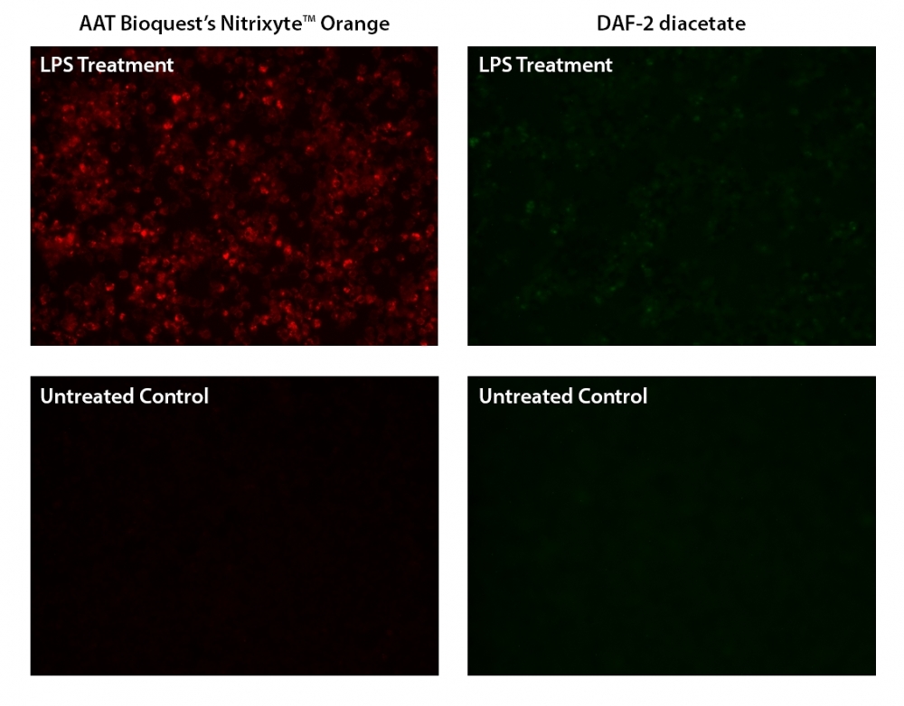
Nitric oxide (NO) is an important biological regulator involved in numbers of physiological and pathological processes. Altered NO production is implicated in various immunological, cardiovascular, neurodegenerative and inflammatory diseases. As a free radical, NO is rapidly oxidized and there is relatively low concentrations of NO existing in vivo. It has been challenging to detect and understand the role of NO in biological systems. Cell Meter™ Fluorimetric Intracellular Nitric Oxide Assay Kit provides a sensitive tool to monitor intracellular NO level in live cells. Nitrixyte™ probes are developed and used in our kit as an excellent replacement for DAF-2 for the detection and imaging of free NO in cells. Compared to the commonly used DAF-2 probe, Nitrixyte™ probes have better photostability and enhanced cell permeability. This particular kit uses Nitrixyte™ Orange that can react with NO to generate a bright orange fluorescent product that has spectral properties similar to Cy3® and TRITC. Nitrixyte™ Orange can be readily loaded into live cells, and its fluorescence signal can be conveniently monitored using the filter set of Cy3® or TRITC. This kit is optimized for fluorescence imaging and microplate reader applications.
Platform
Fluorescence microplate reader
Excitation | 540 nm |
Emission | 590 nm |
Cutoff | 570 nm |
Recommended plate | Black wall/clear bottom |
Instrument specification(s) | Bottom read mode |
Components
Example protocol
AT A GLANCE
Protocol summary
- Prepare cells in growth medium
- Incubate cells with test compounds and Nitrixyte™ Orange working solution at 37oC for a desired period
- Add Assay Buffer II
- Monitor fluorescence intensity (bottom read mode ) at Ex/Em = 540/590 nm (Cutoff = 570 nm) or fluorescence microscope using TRITC filter
Important notes
Thaw all the kit components at room temperature before starting the experiment.
PREPARATION OF WORKING SOLUTION
Add 20 µL of 500X Nitrixyte™ Orange stock solution (Component A) into 10 mL of Assay Buffer I (Component B) and mix well to make Nitrixyte™ Orange working solution. This Nitrixyte™ Orange working solution is stable for at least 2 hours at room temperature. Protect from light. Note: 20 µL of 500X Nitrixyte™ Orange stock solution is enough for one plate.
For guidelines on cell sample preparation, please visit
https://www.aatbio.com/resources/guides/cell-sample-preparation.html
SAMPLE EXPERIMENTAL PROTOCOL
- To stimulate endogenous NO, treat cells with 10 µL of 10X test compounds (96-well plate) or 5 µL of 5X test compounds (384-well plate) in cell culture medium or your desired buffer (such as PBS or HHBS). For control wells (untreated cells), add the corresponding amount of medium or compound buffer. Note: It is not necessary to wash cells before adding compound. However, if tested compounds are serum sensitive, growth medium and serum factors can be aspirated away before adding compounds. Add 90 µL/well (96-well plate) and 20 µL/well (384-well plate) of 1X Hank’s salt solution and 20 mM Hepes buffer (HHBS) or the buffer of your choice after aspiration. Alternatively, cells can be grown in serum-free media.
- Add 100 µL/well (96-well plate) or 25 µL/well (384-well plate) of Nitrixyte™ Orange working solution in the cell plate. Co-incubate cells with test compound and Nitrixyte™ Orange working solution at 37°C for desired period of time, protected from light. Note: DO NOT remove the test compounds. For a NONOate positive control treatment: Cells were incubated with Nitrixyte™ Orange working solution at 37°C for 30 minutes. The working solution was removed and cells were further incubated with 1 mM DEA/NONOate at 37°C for 30 minutes to generate nitric oxide. Note: We have used Raw 264.7 cells incubated with 0.5X Nitrixyte™ Orange, 20 µg/mL of lipopolysaccharide (LPS) and 1 mM L-Arginine (L-Arg) in cell culture medium at 37°C for 16 hours. See Figure 1 for details.
- Remove solution in each well.
- Add Assay Buffer II (Component C) 100 µL/well for a 96-well plate or 25 µL/well for a 384-well plate. Note: DO NOT wash cells before adding Assay Buffer II.
- Monitor the fluorescence increase using microplate reader at Ex/Em = 540/590 nm (Cutoff = 570 nm) with bottom read mode, or take images using fluorescence microscope with a TRITC filter.
Images

Figure 1. Fluorescence images of endogenous nitric oxide (NO) detection in RAW 264.7 macrophage. Cells were incubated with AAT’s Nitrixyte™ Orange (Left) or DAF-2 diacetate (Right) at the same concentration, then treated with or without 20 µg/mL of lipopolysaccharide (LPS) and 1 mM L-arginine (L-Arg) at 37°C for 16 hours. The fluorescence signals were measured using a fluorescence microscope equipped with a TRITC (Left) or FITC (Right) filter set, respectively.

Figure 2. Detection of exogenous nitric oxide (NO) in cells upon DEA/NONOate treatment (NO donor) using Cell Meter™ Fluorimetric Intracellular Nitric Oxide Activity Assay Kit (Cat#16350). CHO-K1 and HeLa cells were incubated with Nitrixyte™ Orange working solution at 37 ºC for 30 minutes. The working solution was removed to stop the staining. The cells were further treated with or without 1mM DEA/NONOate in HBSS with 1 mM HEPES (pH=6.2) buffer at 37 ºC for 30 minutes. The solution in each well was removed, and Assay Buffer II was added before fluorescence measurement. The fluorescence signal was monitored at Ex/Em = 540/590 nm (cut off = 570 nm) with bottom read mode using a FlexStation microplate reader (Molecular Devices).

Figure 3. Microplate reader measurement of exogenous nitric oxide (NO) in HeLa cells upon DEA NONOate treatment (NO donor). Cells were incubated with AAT’s Nitrixyte™ Orange or DAF-2 diacetate at the same concentration for 30 minutes. The cells were then treated with or without 1 mM DEA NONOate at 37°C for 30 minutes. The fluorescence signals were measured with bottom read mode at Ex/Em=540/590 nm or Ex/Em=490/530 nm, respectively. Compared to DAF-2 diacetate, Nitrixyte™ Orange showed a significantly higher signal-to-noise ratio (B) as calculated by dividing RFU of NONOate treated sample by the corresponding untreated control (A).
Citations
View all 4 citations: Citation Explorer
New insights into posttranslational modifications of proteins during bull sperm capacitation
Authors: Mostek-Majewska, Agnieszka and Majewska, Anna and Janta, Anna and Ciereszko, Andrzej
Journal: Cell Communication and Signaling (2023): 1--23
Authors: Mostek-Majewska, Agnieszka and Majewska, Anna and Janta, Anna and Ciereszko, Andrzej
Journal: Cell Communication and Signaling (2023): 1--23
Fluorescent real-time quantitative measurements of intracellular peroxynitrite generation and inhibition
Authors: Luo, Zhen and Zhao, Qin and Liu, Jixiang and Liao, Jinfang and Peng, Ruogu and Xi, Yunting and Diwu, Zhenjun
Journal: Analytical biochemistry (2017): 44--48
Authors: Luo, Zhen and Zhao, Qin and Liu, Jixiang and Liao, Jinfang and Peng, Ruogu and Xi, Yunting and Diwu, Zhenjun
Journal: Analytical biochemistry (2017): 44--48
Inducible nitric oxide synthase (iNOS) is a novel negative regulator of hematopoietic stem/progenitor cell trafficking
Authors: Adamiak, Mateusz and Abdelbaset-Ismail, Ahmed and Moore, Joseph B and Zhao, J and Abdel-Latif, Ahmed and Wysoczynski, Marcin and Ratajczak, Mariusz Z
Journal: Stem cell reviews and reports (2017): 92--103
Authors: Adamiak, Mateusz and Abdelbaset-Ismail, Ahmed and Moore, Joseph B and Zhao, J and Abdel-Latif, Ahmed and Wysoczynski, Marcin and Ratajczak, Mariusz Z
Journal: Stem cell reviews and reports (2017): 92--103
Inducible Nitric Oxide Synthase (iNOS) Is a Novel Negative Regulator of Hematopoietic Stem/Progenitor Cell Trafficking
Authors: Adamiak, Mateusz and Abdelbaset-Ismail, Ahmed and Moore, Joseph B and Zhao, J and Abdel-Latif, Ahmed and Wysoczynski, Marcin and Ratajczak, Mariusz Z
Journal: Stem Cell Reviews and Reports (2016): 1--12
Authors: Adamiak, Mateusz and Abdelbaset-Ismail, Ahmed and Moore, Joseph B and Zhao, J and Abdel-Latif, Ahmed and Wysoczynski, Marcin and Ratajczak, Mariusz Z
Journal: Stem Cell Reviews and Reports (2016): 1--12
References
View all 139 references: Citation Explorer
Pitfalls and limitations in using 4,5-diaminofluorescein for evaluating the influence of polyphenols on nitric oxide release from endothelial cells
Authors: Uhlenhut K, Hogger P.
Journal: Free Radic Biol Med (2012): 2266
Authors: Uhlenhut K, Hogger P.
Journal: Free Radic Biol Med (2012): 2266
Effects of moderate electrical stimulation on reactive species production by primary rat skeletal muscle cells: cross talk between superoxide and nitric oxide production
Authors: Lambertucci RH, Silveira Ldos R, Hirabara SM, Curi R, Sweeney G, Pithon-Curi TC.
Journal: J Cell Physiol (2012): 2511
Authors: Lambertucci RH, Silveira Ldos R, Hirabara SM, Curi R, Sweeney G, Pithon-Curi TC.
Journal: J Cell Physiol (2012): 2511
Improved measurements of intracellular nitric oxide in intact microvessels using 4,5-diaminofluorescein diacetate
Authors: Zhou X, He P.
Journal: Am J Physiol Heart Circ Physiol (2011): H108
Authors: Zhou X, He P.
Journal: Am J Physiol Heart Circ Physiol (2011): H108
Aging negatively affects estrogens-mediated effects on nitric oxide bioavailability by shifting ERalpha/ERbeta balance in female mice
Authors: Novensa L, Novella S, Medina P, Segarra G, Castillo N, Heras M, Hermenegildo C, Dantas AP.
Journal: PLoS One (2011): e25335
Authors: Novensa L, Novella S, Medina P, Segarra G, Castillo N, Heras M, Hermenegildo C, Dantas AP.
Journal: PLoS One (2011): e25335
Temporal and spatial correlation of platelet-activating factor-induced increases in endothelial [Ca(2)(+)]i, nitric oxide, and gap formation in intact venules
Authors: Zhou X, He P.
Journal: Am J Physiol Heart Circ Physiol (2011): H1788
Authors: Zhou X, He P.
Journal: Am J Physiol Heart Circ Physiol (2011): H1788
Polyamines, polyamine oxidases and nitric oxide in development, abiotic and biotic stresses
Authors: Wimalasekera R, Tebartz F, Scherer GF.
Journal: Plant Sci (2011): 593
Authors: Wimalasekera R, Tebartz F, Scherer GF.
Journal: Plant Sci (2011): 593
Rapid upregulation of cytoprotective nitric oxide in breast tumor cells subjected to a photodynamic therapy-like oxidative challenge
Authors: Bhowmick R, Girotti AW.
Journal: Photochem Photobiol (2011): 378
Authors: Bhowmick R, Girotti AW.
Journal: Photochem Photobiol (2011): 378
Sleep deprivation triggers inducible nitric oxide-dependent nitric oxide production in wake-active basal forebrain neurons
Authors: Kalinchuk AV, McCarley RW, Porkka-Heiskanen T, Basheer R.
Journal: J Neurosci (2010): 13254
Authors: Kalinchuk AV, McCarley RW, Porkka-Heiskanen T, Basheer R.
Journal: J Neurosci (2010): 13254
Production and scavenging of nitric oxide by barley root mitochondria
Authors: Gupta KJ, Kaiser WM.
Journal: Plant Cell Physiol (2010): 576
Authors: Gupta KJ, Kaiser WM.
Journal: Plant Cell Physiol (2010): 576
Production of Nitric Oxide within the Aplysia Californica Nervous System
Authors: Ye X, Xie F, Romanova EV, Rubakhin SS, Sweedler JV.
Journal: ACS Chem Neurosci (2010): 182
Authors: Ye X, Xie F, Romanova EV, Rubakhin SS, Sweedler JV.
Journal: ACS Chem Neurosci (2010): 182
Application notes
A Novel Fluorescent Probe for Imaging and Detecting Hydroxyl Radical in Living Cells
Abbreviation of Common Chemical Compounds Related to Peptides
A Comparison of Fluorescent Red Calcium Indicators for Detecting Intracellular Calcium Mobilization in CHO Cells
A Meta-Analysis of Common Calcium Indicators
A New Red Fluorescent & Robust Screen Quest™ Rhod-4™ Ca2+Indicator for Screening GPCR & Ca2+ Channel Targets