logo
AAT Bioquest

Cell Meter™ Fluorimetric Intracellular pH Assay Kit

Intracellular pH change are implicated in diverse physiological and pathological processes, including cell proliferation, apoptosis, fertilization, malignancy, multidrug resistance, ion transport, lysosomal storage disorders and Alzheimer's disease. The Cell Meter™ Fluorimetric Intracellular pH Assay Kit utilizes AAT Bioquest's proprietary fluorescent indicator for measuring the relative intracellular pH changes. It is a homogeneous, kinetic, live-cell fluorescent assay that utilizes either a standard procedure or acid-load procedure. The standard protocol is designed for measuring the therapeutic targets of interest with a decrease in intracellular pH upon treatment. The 'Acid-Load' procedure is designed to measure the increase of intracellular pH associated with changes in cellular metabolism due to GPCR activation or growth factor activity. With the 'Acid-Load' procedure ammonium chloride solution is added after the fluorescent pH dye is loaded into cells in a minimum volume. This 'acid-loading' step is followed by the addition of agonist in a relatively large volume (~4X) of buffer. The sudden volume change initiates an efflux of ammonia (NH3) from the cells causing a rapid decrease in intracellular pH, and thus a decrease in fluorescence signal. The effect of agonist on the subsequent recovery of intracellular pH is measured by the relative fluorescence signal increase.
Example protocol

AT A GLANCE

Protocol Summary
for Standard Cell Load (One Plate)
  1. Prepare cells in growth medium
  2. Add equal volume of RatioWorks™ BCFL, AM dye-working solution
  3. Incubate at 37 °C for 1 hour
  4. Read Fluorescence at Ex/Em= 490/535 nm (Cutoff = 515 nm) with 50 µL/well compound addition (or 500/530 nm and 440/530 nm for ratio) 

Protocol Summary
for Acid-Load (One 96-well Plate)
  1. Prepare cells in growth medium
  2. Remove the growth medium
  3. Add RatioWorks™ BCFL, AM dye-working solution (50 µL/well/96-well plate)
  4. Incubate at 37°C for 1 hour
  5. Add 220 mM NH4Cl (5 µL/well)
  6. Incubate at RT for 15 minutes
  7. Read Fluorescence at Ex/Em= 490/535 nm (Cutoff = 515 nm) with 200 µL/well compound addition (or 500/530 nm and 440/530 nm for ratio) 
Important      Thaw all the kit components at room temperature before starting the experiment.

CELL PREPARATION

For guidelines on cell sample preparation, please visit https://www.aatbio.com/resources/guides/cell-sample-preparation.html

PREPARATION OF STOCK SOLUTIONS

Unless otherwise noted, all unused stock solutions should be divided into single-use aliquots and stored at -20 °C after preparation. Avoid repeated freeze-thaw cycles.

RatioWorks™ BCFL, AM stock solution
Add 200 µL DMSO into the vial of RatioWorks™ BCFL, AM (Component A) and mix well to make RatioWorks™ BCFL, AM stock solution.
Note      20 µL of reconstituted RatioWorks™ BCFL, AM stock solution is enough for 1 plate of Standard Cell Load. 10 µL of reconstituted RatioWorks™ BCFL, AM stock solution is enough for 1 plate of Acid Load. RatioWorks™ BCFL, AM stock solution can be stored at ≤ -20 oC for one month if the tubes are sealed tightly. Protect from light.

PREPARATION OF WORKING SOLUTION

1. For Standard Cell Load (One Plate)
  1. Add 1 mL of 10X Pluronic F127 Plus (Component B) into 9 mL of HHBS (Component C) and mix well to make 1X assay buffer.
    Note      For cells that require probenecid for loading (e.g. CHO cells), dilute 50 mM ReadiUse™ Probenecid (Component D) at concentration of 1 to 5 mM (prefer 5 mM for CHO cells).
  2. Add 20 µL of reconstituted RatioWorks™ BCFL, AM stock solution into 10 mL of 1X assay buffer and mix well to make RatioWorks™ BCFL, AM dye-working solution. This RatioWorks™ BCFL, AM dye-working solution is stable for at least 2 hours at room temperature. 

2. For Acid-Load (One 96-well Plate)
  1. Add 1 mL of 10X Pluronic F127 Plus (Component B) into 4 mL of HHBS (Component C) and mix well to make 1X assay buffer.
    Note      For cells that require probenecid for loading (e.g. CHO cells), dilute 50 mM ReadiUse™ Probenecid (Component D) at concentration of 0.5 to 2.5 mM (prefer 2.5 mM for CHO cells).
  2. Add 10 µL of reconstituted RatioWorks™ BCFL, AM stock solution into 5 mL of 1X assay buffer and mix well to make RatioWorks™ BCFL, AM dye-working solution. This RatioWorks™ BCFL, AM dye-working solution is stable for at least 2 hours at room temperature. 

SAMPLE EXPERIMENTAL PROTOCOL

Run pH Assay for Standard Cell Load
  1. Add 100 µL/well (96-well plate) or 25 µL/well (384-well plate) RatioWorks™ BCFL, AM dye-working solution into the cell plate.
    Note      It is important to replace the growth medium with HHBS buffer (100 µL/well for 96-well plate or 25 µL/well for 384-well plate before dye-loading) if your compounds interfere with the serum.
  2. Incubate the dye-loading plate in cell incubator for 30 minutes, and then incubate the plate at room temperature for another 30 minutes.
    Note      If the assay requires 37°C, perform the experiment immediately without further room temperature incubation.
  3. Prepare the compound plates by using HHBS or your desired buffer.
  4. Run the pH assay by monitoring the fluorescence at Ex/Em = 490/535 nm (Cutoff = 515 nm) or 500/530 nm and 440/530 nm (Cutoff = 515 nm) for ratio measurements. The compound addition is 50 µL/well (96-well plate) or 25 µL/well (384-well plate). 
    Note      The assay should be complete within 3 to 5 min after compound addition, however a minimum of 8 min data collection are recommended for during assay development. 

Run pH Assay for Acid-Load
  1. Remove the growth medium from the cell plate.
  2. Add 50 µL/well/96-well plate RatioWorks™ BCFL, AM dye-working solution into the cell plate.
  3. Incubate the dye-loading plate in cell incubator for 30 minutes, and then incubate the plate at room temperature for another 30 minutes. Note: If the assay requires 37°C, perform the experiment immediately without further room temperature incubation.
  4. Add 5 µL of 220 mM NH4Cl and centrifuge the plates for 5 seconds. Incubate 15 minutes at room temperature.
    Note      NH4Cl solution should be prepared freshly in HHBS (Component C).
  5. Prepare the compound plates by using HHBS or your desired buffer.
  6. Run the pH assay by monitoring the fluorescence at Ex/Em = 490/535 nm (Cutoff = 515 nm) or 500/530 nm and 440/530 nm (Cutoff = 515 nm) for ratio measurements. The compound addition is 200 µL/well/96-well plate.
    Note      The assay should be complete within 3 to 5 min after compound addition, however a minimum of 8 min data collection are recommended for during assay development. 

Citations

View all 2 citations: Citation Explorer
Empagliflozin suppressed cardiac fibrogenesis through sodium-hydrogen exchanger inhibition and modulation of the calcium homeostasis
Authors: Chung, Cheng-Chih and Lin, Yung-Kuo and Chen, Yao-Chang and Kao, Yu-Hsun and Yeh, Yung-Hsin and Trang, Nguyen Ngoc and Chen, Yi-Jen
Journal: Cardiovascular Diabetology (2023): 1--15
The Redox Status of Cancer Cells Supports Mechanisms behind the Warburg Effect
Authors: Moreira, Jorgelindo Da Veiga and Hamraz, Minoo and Abolhassani, Mohammad and Bigan, Erwan and Pérès, Sabine and Paulevé, Loic and Nogueira, Marcel Levy and Steyaert, Jean-Marc and Schwartz, Laurent
Journal: Metabolites (2016): 33

References

View all 34 references: Citation Explorer
Determination of intracellular pH using sensitive, clickable fluorescent probes
Authors: Yapici NB, M and alapu SR, Chew TL, Khuon S, Bi L.
Journal: Bioorg Med Chem Lett (2012): 2440
A two-step pH-dependent liquid-liquid extraction combined with HPLC-fluorescence method for the determination of 10-hydroxycamptothecin in mouse liver tissue
Authors: Zheng J, Guo H, Guo N, Ma W, Jing L, Zhang R, Dai Z, Yan X, Wang Y, Wang Z.
Journal: Pharm Biol (2012): 954
Aqueous synthesis of CdTe/CdSe core/shell quantum dots as pH-sensitive fluorescence probe for the determination of ascorbic acid
Authors: Yang SS, Ren CL, Zhang ZY, Hao JJ, Hu Q, Chen XG.
Journal: J Fluoresc (2011): 1123
Flow injection small-volume fiber-optic pH sensor based on evanescent wave excitation and fluorescence determination
Authors: Xiong Y, Huang Y, Ye Z, Guan Y.
Journal: J Fluoresc (2011): 1137
Isoelectric point determination for Glossoscolex paulistus extracellular hemoglobin: oligomeric stability in acidic pH and relevance to protein-surfactant interactions
Authors: Santiago PS, Carvalho FA, Domingues MM, Carvalho JW, Santos NC, Tabak M.
Journal: Langmuir (2010): 9794
Page updated on October 23, 2024

Ordering information

Price
Unit size
Catalog Number21180
Quantity
Add to cart

Additional ordering information

Telephone1-800-990-8053
Fax1-800-609-2943
Emailsales@aatbio.com
InternationalSee distributors
Bulk requestInquire
Custom sizeInquire
Technical SupportContact us
Purchase orderSend to sales@aatbio.com
ShippingStandard overnight for United States, inquire for international
Request quotation

Storage, safety and handling

H-phraseH303, H313, H333
Hazard symbolXN
Intended useResearch Use Only (RUO)
R-phraseR20, R21, R22
UNSPSC12352200

Platform

Fluorescence microplate reader

Excitation490 nm
Emission535 nm
Cutoff515 nm
Recommended plateBlack well, Clear bottom

Other instruments

ArrayScan, FDSS, FLIPR, FlexStation, IN Cell Analyzer, NOVOStar, ViewLux

Components

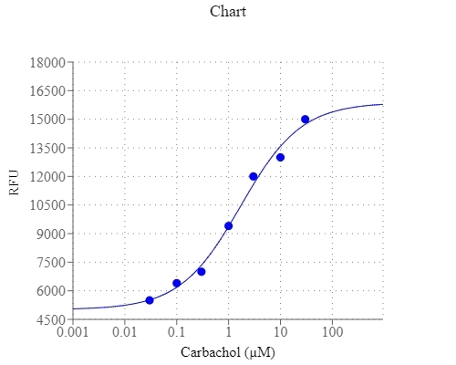
Carbachol dose response in CHO-M1 cell. CHO-M1 cells were seeded overnight in 60,000 cells per 100 µL per well in a 96- well black wall/clear bottom costar plate. The growth medium was replaced with 50 µL/well of RatioWorks™ BCFL, AM dye-loading solution for 37°C for 1 hour, follow by 15 minutes incubation with 5  µL/well of 220 mM NH4Cl. Carbachol (200µL/well) was added by FlexStation (Molecular Devices) to achieve the final indicated concentrations. The fluorescent signal was generated using Ex/Em = 490/535 nm (Cutoff = 515 nm).
Effects of empagliflozin on Na+/H+ exchanger (NHE) and downstream signaling. Averaged data of the intracellular pH in the control cells and empagliflozin (1 μmol/L)-treated atrial fibroblasts cotreated with or without cariporide (10 μmol/L) for 48 h (n = 6 experiments, statistic test by one-way repeated measures ANOVA). Source: Graph from <b>Empagliflozin suppressed cardiac fibrogenesis through sodium-hydrogen exchanger inhibition and modulation of the calcium homeostasis</b> by Chung et al., <em>Cardiovascular Diabetology</em>, Feb. 2023.