Products
Services
Resources
Selection Guides
About
Cell Meter™ Fluorimetric Mitochondrial Superoxide Activity Assay Kit
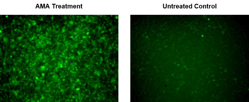
Green Fluorescence
Mitochondria are major producers of cellular superoxide. The production of low to moderate levels of superoxide is critical for the proper regulation of many essential cellular processes including gene expression, signal transduction, and muscle adaptation to endurance exercise training. Uncontrolled mitochondrial superoxide production can trigger cellular oxidative damage that contributes to the pathogenesis of a wide variety of disorders including cancer, cardiovascular diseases, neurodegenerative diseases and aging. The detection of intracellular mitochondrial superoxide is of great importance to understanding proper cellular redox regulation and the impact of its dysregulation on various pathologies. Cell Meter™ Fluorimetric Intracellular Superoxide Detection Kit uses MitoROS™ 520, our unique superoxide indicator, to quantify superoxide level in live cells. MitoROS™ 520 is cell permeant and can rapidly and selectively detect superoxide in mitochondria. It generates green fluorescence upon reacting with superoxide. The Cell Meter™ Fluorimetric Mitochondrial Superoxide Activity Assay Kit provides a sensitive, one-step fluorimetric assay to detect mitochondrial superoxide in live cells with one hour incubation. This kit can be used for flow cytometry and fluorescence microscopy applications.
| Catalog | Size | Price | Quantity |
|---|---|---|---|
| 16060 | 200 Tests | Price |

Spectral properties
| Excitation (nm) | 513 |
| Emission (nm) | 537 |
Usage and storage
| Intended use | Research Use Only (RUO) |
Instrument settings
| Flow cytometer | |
| Excitation | 488 nm laser |
| Emission | 530/30 nm filter |
| Instrument specification(s) | FITC channel |
| Fluorescence microscope | |
| Excitation | FITC filter set |
| Emission | FITC filter set |
| Recommended plate | Black wall/clear bottom |
| Instrument specification(s) | FITC filter set |
Contact us
| Telephone | |
| Fax | |
| sales@aatbio.com | |
| International | See distributors |
| Bulk request | Inquire |
| Custom size | Inquire |
| Technical Support | Contact us |
| Request quotation | Request |
| Purchase order | Send to sales@aatbio.com |
| Shipping | Standard overnight for United States, inquire for international |
Page updated on July 16, 2026