logo
AAT Bioquest

Cell Meter™ Fluorimetric Mitochondrial Superoxide Activity Assay Kit *Green Fluorescence*

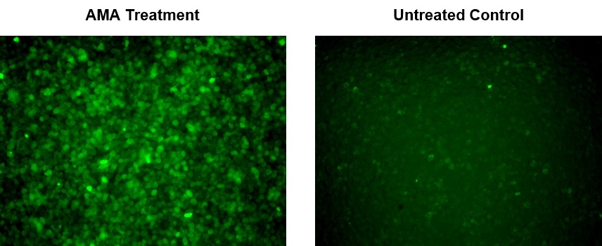
Fluorescence images of superoxide measurement in macrophage cells using cat#16060. RAW 264.7 cells at 100,000 cells/well/100 µL were seeded overnight in a 96-well black wall/clear bottom plate. AMA Treatment: Cells were treated with 5 µM Antimycin A (AMA) at 37 °C for 2 hours, then incubated with MitoROS™ 520 for 1 hour. Untreated Control: RAW 264.7 cells were incubated with MitoROS™ 520 at 37 °C for 1 hour without AMA treatment. The fluorescence signal was measured using fluorescence microscope with a FITC filter
Fluorescence images of endogenous superoxide measurement in macrophage cells using Cat#16060. RAW 264.7 cells at 100,000 cells/well/100 µL were seeded overnight in a 96-well black wall/clear bottom plate. LPS Treatment: Cells were incubated with MitoROS™ 520 for 1 hour, then treated with 200 μg/mL of lipopolysaccharide (LPS) at 37 ºC for 16 hours. Untreated Control: RAW 264.7 cells were incubated with MitoROS™ 520 at 37 ºC for 1 hour without LPS treatment. The fluorescence signal was measured using fluorescence microscope with a FITC filter.
Ordering information
Price
Catalog Number
Unit Size
Quantity
Add to cart
Additional ordering information
Telephone1-800-990-8053
Fax1-800-609-2943
Emailsales@aatbio.com
InternationalSee distributors
Bulk requestInquire
Custom sizeInquire
ShippingStandard overnight for United States, inquire for international
Request quotation
Spectral properties
Excitation (nm)513
Emission (nm)537
Storage, safety and handling
Certificate of OriginDownload PDF
H-phraseH303, H313, H333
Hazard symbolXN
Intended useResearch Use Only (RUO)
R-phraseR20, R21, R22
UNSPSC12352200
Related products
Cell Meter™ Intracellular Fluorimetric Hydrogen Peroxide Assay Kit *Green Fluorescence*
Cell Meter™ Intracellular Fluorimetric Hydrogen Peroxide Assay Kit *Blue Fluorescence*
Cell Meter™ Intracellular Fluorimetric Hydrogen Peroxide Assay Kit *Blue Fluorescence Optimized for Flow Cytometry*
Cell Meter™ Intracellular Fluorimetric Hydrogen Peroxide Assay Kit *Green Fluorescence Optimized for Flow Cytometry*
Cell Meter™ Mitochondrial Hydroxyl Radical Detection Kit *Red Fluorescence*
Cell Meter™ Fluorimetric Intracellular Peroxynitrite Assay Kit *Green Fluorescence*
Cell Meter™ Fluorimetric Intracellular Peroxynitrite Assay Kit *Optimized for Flow Cytometry*
Cell Meter™ Fluorimetric Intracellular Nitric Oxide (NO) Activity Assay Kit *Orange Fluorescence Optimized for Microplate Reader*
Cell Meter™ Fluorimetric Intracellular Nitric Oxide (NO) Activity Assay Kit *Orange Fluorescence Optimized for Flow Cytometry*
Cell Meter™ Fluorimetric Intracellular Nitric Oxide (NO) Activity Assay Kit *Red Fluorescence Optimized for Flow Cytometry*
Cell Meter™ Live Cell Caspase 3/7 Binding Assay Kit *Green Fluorescence*
Cell Meter™ Live Cell Caspase 3/7 Binding Assay Kit *Red Fluorescence*
Cell Meter™ Live Cell Caspase 1 Binding Assay Kit *Green Fluorescence*
Cell Meter™ Live Cell Caspase 2 Binding Assay Kit *Green Fluorescence*
Cell Meter™ Live Cell Caspase 6 Binding Assay Kit *Green Fluorescence*
Cell Meter™ Live Cell Caspase 8 Binding Assay Kit *Green Fluorescence*
Cell Meter™ Live Cell Caspase 9 Binding Assay Kit *Green Fluorescence*
Cell Meter™ Live Cell Caspase 10 Binding Assay Kit *Green Fluorescence*
Cell Meter™ Live Cell Caspase 13 Binding Assay Kit *Green Fluorescence*
Cell Meter™ No-Wash Live Cell Caspase 3/7 Activity Assay Kit *Blue Fluorescence*
Cell Meter™ No-Wash Live Cell Caspase 3/7 Activity Assay Kit *Red Fluorescence*
Cell Meter™ Fluorimetric Intracellular pH Assay Kit
Cell Meter™ Fluorimetric Cell Cytotoxicity Assay Kit
Cell Meter™ Cell Viability Assay Kit *Red Fluorescence*
Cell Meter™ Cell Viability Assay Kit *Blue Fluorescence with 405 nm Excitation*
Cell Meter™ Cell Viability Assay Kit *Blue Fluorescence*
Cell Meter™ Cell Viability Assay Kit *Green Fluorescence*
Cell Meter™ Cell Viability Assay Kit *NIR Fluorescence Optimized for Fluorescence Microplate Reader*
Cell Meter™ Phosphatidylserine Apoptosis Assay Kit *Blue Fluorescence Optimized for Microplate Readers*
Cell Meter™ Phosphatidylserine Apoptosis Assay Kit *Green Fluorescence Optimized for Microplate Readers*
Cell Meter™ Phosphatidylserine Apoptosis Assay Kit *Red Fluorescence Optimized for Microplate Readers*
Cell Meter™ Phosphatidylserine Apoptosis Assay Kit *Deep Red Fluorescence Optimized for Microplate Readers*
Cell Meter™ Phosphatidylserine Apoptosis Assay Kit *Orange Fluorescence Optimized for Microplate Readers*
Cell Meter™ Caspase 3/7 Activity Apoptosis Assay Kit *Blue Fluorescence*
Cell Meter™ Caspase 3/7 Activity Apoptosis Assay Kit *Green Fluorescence*
Cell Meter™ Caspase 3/7 Activity Apoptosis Assay Kit *Red Fluorescence*
Cell Meter™ Caspase 8 Activity Apoptosis Assay Kit *Green Fluorescence*
Cell Meter™ Caspase 9 Activity Apoptosis Assay Kit *Green Fluorescence*
Cell Meter™ JC-10 Mitochondrion Membrane Potential Assay Kit *Optimized for Microplate Assays*
Cell Meter™ JC-10 Mitochondrion Membrane Potential Assay Kit *Optimized for Flow Cytometry Assays*
Cell Meter™ NIR Mitochondrion Membrane Potential Assay Kit *Optimized for Flow Cytometry*
Cell Meter™ NIR Mitochondrion Membrane Potential Assay Kit *Optimized for Microplate Reader*
Cell Meter™ Mitochondrion Membrane Potential Assay Kit *Orange Fluorescence Optimized for Flow Cytometry*
Cell Meter™ Mitochondrion Membrane Potential Assay Kit *Orange Fluorescence Optimized for Microplate Reader*
Cell Meter™ Intracellular GSH Assay Kit *Optimized for Flow Cytometry*
Cell Meter™ Nuclear Apoptosis Assay Kit *Green Fluorescence Optimized for Flow Cytometry*
Cell Meter™ Caspase 8 Activity Apoptosis Assay Kit *Blue Fluorescence*
Cell Meter™ Caspase 9 Activity Apoptosis Assay Kit *Blue Fluorescence*
Cell Meter™ Caspase 8 Activity Apoptosis Assay Kit *Red Fluorescence*
Cell Meter™ Caspase 9 Activity Apoptosis Assay Kit *Red Fluorescence*
Cell Meter™ Multiplexing Caspase 3/7, 8 and 9 Activity Assay Kit *Triple Fluorescence Colors*
Cell Meter™ Generic Fluorimetric Caspase Activity Assay Kit *Green Fluorescence Optimized for Flow Cytometry*
Cell Meter™ Generic Fluorimetric Caspase Binding Assay Kit *Red Fluorescence Optimized for Flow Cytometry*
Cell Meter™ Caspase 3/7 Activity Apoptosis Assay Kit *Green Fluorescence Optimized for Flow Cytometry*
Cell Meter™ Annexin V Binding Apoptosis Assay Kit *Green Fluorescence Optimized for Flow Cytometry*
Cell Meter™ Annexin V Binding Apoptosis Assay Kit *Orange Fluorescence Optimized for Flow Cytometry*
Cell Meter™ Annexin V Binding Apoptosis Assay Kit *Red Fluorescence Optimized for Flow Cytometry*
Cell Meter™ Annexin V Binding Apoptosis Assay Kit *Deep Red Fluorescence Optimized for Flow Cytometry*
Cell Meter™ Annexin V Binding Apoptosis Assay Kit *Blue Fluorescence Excited at 405 nm*
Cell Meter™ Annexin V Binding Apoptosis Assay Kit *Green Fluorescence Excited at 405 nm*
Cell Meter™ Annexin V Binding Apoptosis Assay Kit *Orange Fluorescence Excited at 405 nm*
Cell Meter™ Phosphatidylserine Apoptosis Assay Kit *Green Fluorescence Optimized for Flow Cytometry*
Cell Meter™ Phosphatidylserine Apoptosis Assay Kit *Deep Red Fluorescence Optimized for Flow Cytometry*
Cell Meter™ Phosphatidylserine Apoptosis Assay Kit *Blue Fluorescence Excited at 405 nm*
Cell Meter™ Phosphatidylserine Apoptosis Assay Kit *Green Fluorescence Excited at 405 nm*
Cell Meter™ APC-Annexin V Binding Apoptosis Assay Kit *Optimized for Flow Cytometry*
Cell Meter™ PE-Annexin V Binding Apoptosis Assay Kit *Optimized for Flow Cytometry*
Cell Meter™ Apoptotic and Necrotic Multiplexing Detection Kit I *Triple Fluorescence Colors*
Cell Meter™ Fluorimetric Live Cell Cycle Assay Kit *Green Fluorescence Optimized for Flow Cytometry*
Cell Meter™ Fluorimetric Fixed Cell Cycle Assay Kit *Red Fluorescence Optimized for Flow Cytometry*
Cell Meter™ Apoptotic and Necrotic Multiplexing Detection Kit II *Triple Fluorescence Colors*
Cell Meter™ Live Cell TUNEL Apoptosis Assay Kit *Red Fluorescence*
Cell Meter™ Fluorimetric Live Cell Cycle Assay Kit *Optimized for 405 nm Violet Laser Excitation*
Cell Meter™ Live Cell Caspase 3/7 and Phosphatidylserine Detection Kit *Triple Fluorescence Colors*
Cell Meter™ Fluorimetric Intracellular Total ROS Activity Assay Kit*Green Fluorescence*
Cell Meter™ Fluorimetric Intracellular Total ROS Activity Assay Kit*Red Fluorescence*
Cell Meter™ Fluorimetric Intracellular Total ROS Activity Assay Kit*Orange Fluorescence*
Cell Meter™ Fluorimetric Intracellular Total ROS Activity Assay Kit*Deep Red Fluorescence*
Cell Meter™ Fluorimetric Intracellular Total ROS Activity Assay Kit*Optimized for Flow Cytometry*
Cell Meter™ Autophagy Assay Kit *Blue Fluorescence*
Cell Meter™ Autophagy Fluorescence Imaging Kit
Cell Meter™ Autophagy Assay Kit *Green Fluorescence*
Cell Meter™ FITC-Annexin V Binding Apoptosis Assay Kit *Optimized for Flow Cytometry*
Cell Meter™ Fluorimetric Intracellular Nitric Oxide (NO) Activity Assay Kit *NIR Fluorescence Optimized for Microplate Reader*
Cell Meter™ Fluorimetric Intracellular Nitric Oxide (NO) Activity Assay Kit *NIR Fluorescence Optimized for Flow Cytometry*
Cell Meter™ Colorimetric WST-8 Cell Quantification Kit
Cell Meter™ Multiplexing Live, Apoptotic and Necrotic Cell Detection Kit III *Triple Fluorescence Colors*
Cell Meter™ 2-NBDG Glucose Uptake Assay Kit
Cell Meter™ Intracellular NADH/NADPH Fluorescence Imaging Kit *Red Fluorescence*
Cell Meter™ Intracellular NADH/NADPH Flow Cytometric Analysis Kit *Red Fluorescence*
Cell Meter™ Live Cell TUNEL Apoptosis Assay Kit *Green Fluorescence*
Cell Meter™ No Wash and Probenecid-Free Endpoint Calcium Assay Kit *Optimized for microplate reader*
Cell Meter™ Mitochondrion Membrane Potential Assay Kit *Red Fluorescence Optimized for Flow Cytometry*
Cell Meter™ Mitochondrion Membrane Potential Assay Kit *Red Fluorescence Optimized for Microplate Reader*
Cell Meter™ Intracellular Colorimetric Lipid Peroxidation (MDA) Assay Kit
Cell Meter™ Flow Cytometric Calcium Assay Kit
Cell Meter™ Fluorimetric Live Cell Cycle Assay Kit *Red Fluorescence Optimized for Flow Cytometry*
Cell Meter™ Fluorimetric Cellular Lipid Peroxidation Assay Kit
Cell Meter™ Intracellular NADH/NADPH Fluorescence Imaging Kit *Deep Red Fluorescence*
Cell Meter™ Intracellular NADH/NADPH Flow Cytometric Analysis Kit *Deep Red Fluorescence*
Cell Meter™ Intracellular GSH Assay Kit *Optimized for Flow Cytometry with 405 nm excitation*
Cell Meter™ Mitochondrial Autophagy Imaging Kit *Red Fluorescence*
Cell Meter™ BX520 fixable viability dye
Cell Meter™ BX590 fixable viability dye
Cell Meter™ BX650 fixable viability dye
Cell Meter™ IX830 fixable viability dye
Cell Meter™ RX660 fixable viability dye
Cell Meter™ RX700 fixable viability dye
Cell Meter™ RX780 fixable viability dye
Cell Meter™ VX450 fixable viability dye
Cell Meter™ VX500 fixable viability dye
Cell Meter™ Live Cell Caspase 8 Binding Assay Kit *Red Fluorescence*
Cell Meter™ Fixed Cell and Tissue TUNEL Apoptosis Assay Kit *Green Fluorescence*
Cell Meter™ Fixed Cell and Tissue TUNEL Apoptosis Assay Kit *Red Fluorescence*
Cell Meter™ Fluorimetric Cellular Voltage Assay Kit
Cell Meter™ Cell Adhesion Assay Kit
Cell Meter™ Fixed Cell and Tissue TUNEL Apoptosis Assay Kit *Deep Red Fluorescence*
Cell Meter™ Fixed Cell and Tissue TUNEL Apoptosis Assay Kit *Blue Fluorescence*
Cell Meter™ Fluorescence Gap Junction Tracing Kit
Cell Meter™ Colorimetric MTT Cell Proliferation Kit
Cell Meter™ Live Cell Caspase 3/7 Imaging Kit *Green Fluorescence*
Cell Meter™ Beta-Arrestin Translocation GPCR Signaling Kit
Cell Meter™ Live Cell ATP Assay Kit
Cell Meter™ Colorimetric Cell Cytotoxicity Assay Kit
Cell Meter™ Fluorimetric Phagocytosis Assay Kit *Red Fluorescence*
Cell Meter™ Cellular Senescence Activity Assay Kit *Green Fluorescence*
Cell Meter™ Cellular Senescence Activity Assay Kit *Red Fluorescence*
Cell Meter™ Cell Proliferation Assay Kit
Cell Meter™ Glucose Uptake Imaging Kit *Red Fluorescence*
Cell Meter™ Colorimetric MTS Cell Proliferation Kit
Show More (120)

OverviewpdfSDSpdfProtocol


Excitation (nm)
513
Emission (nm)
537
Mitochondria are major producers of cellular superoxide. The production of low to moderate levels of superoxide is critical for the proper regulation of many essential cellular processes including gene expression, signal transduction, and muscle adaptation to endurance exercise training. Uncontrolled mitochondrial superoxide production can trigger cellular oxidative damage that contributes to the pathogenesis of a wide variety of disorders including cancer, cardiovascular diseases, neurodegenerative diseases and aging. The detection of intracellular mitochondrial superoxide is of great importance to understanding proper cellular redox regulation and the impact of its dysregulation on various pathologies. Cell Meter™ Fluorimetric Intracellular Superoxide Detection Kit uses MitoROS™ 520, our unique superoxide indicator, to quantify superoxide level in live cells. MitoROS™ 520 is cell permeant and can rapidly and selectively detect superoxide in mitochondria. It generates green fluorescence upon reacting with superoxide. The Cell Meter™ Fluorimetric Mitochondrial Superoxide Activity Assay Kit provides a sensitive, one-step fluorimetric assay to detect mitochondrial superoxide in live cells with one hour incubation. This kit can be used for flow cytometry and fluorescence microscopy applications.
Platform


Flow cytometer

Excitation488 nm laser
Emission530/30 nm filter
Instrument specification(s)FITC channel

Fluorescence microscope

ExcitationFITC filter set
EmissionFITC filter set
Recommended plateBlack wall/clear bottom
Instrument specification(s)FITC filter set

Components


Example protocol


AT A GLANCE

Protocol summary (Fluorescence Microscope)

  1. Prepare cells in growth medium
  2. Treat the cells with test compounds to induce superoxide
  3. Add MitoROS™ 520 working solution
  4. Incubate the cells at 37°C for 1 hour
  5. Monitor the fluorescence using FITC fliter set

Protocol summary (Flow Cytometry)

  1. Prepare cells in growth medium
  2. Treat the cells with test compounds to induce superoxide
  3. Add MitoROS™ 520 stock solution and incubate the cells at 37°C for 1 hour
  4. Monitor the fluorescence intensity with a flow cytometer using 530/30 nm filter (FITC channel)

Important notes
Thaw all the components at room temperature before use.

PREPARATION OF STOCK SOLUTION

Unless otherwise noted, all unused stock solutions should be divided into single-use aliquots and stored at -20 °C after preparation. Avoid repeated freeze-thaw cycles.

1. MitoROS™ 520 stock solution (500X):
Add 50 µL of DMSO (Component C) into the vial of MitoROS™ 520 (Component A) and mix well. Note: 25 µL of reconstituted MitoROS™ 520 stock solution is enough for 1 plate. Note: Unused portion can be aliquoted and stored at < -20 °C for more than one month if the tubes are sealed tightly and kept from light. Avoid repeated freeze-thaw cycles.

PREPARATION OF WORKING SOLUTION

Only for Fluorescence Microscope
Add 5 μL of 500X DMSO reconstituted MitoROS™ 520 stock solution into 2 mL of Assay Buffer (Component B) and mix well. Note: This working solution is not stable and needs to be prepared freshly before use.

For guidelines on cell sample preparation, please visit
https://www.aatbio.com/resources/guides/cell-sample-preparation.html

SAMPLE EXPERIMENTAL PROTOCOL

For Fluorescence Microscopes/96-Well Microplates:

  1. Treat cells with 10 µL of 10X test compounds (96-well plate) or 5 µL of 5X test compounds (384-well plate) in medium or your desired buffer (such as PBS or HHBS). For control wells (untreated cells), add the corresponding amount of compound buffer.

  2. To induce superoxide, incubate the cell at 37°C for a desired period of time, protected from light. Note: We treated RAW 264.7 macrophage cells with 5 µM Antimycin A (AMA) at 37°C for 2 hours to induce superoxide. See Figure 1 for details.

  3. Add 100 µL/well (96-well plate) or 25 µL/well (384-well plate) of MitoROS™ 520 working solution into the cell plate.

  4. Incubate the cells at 37°C for 1 hour, and take images using fluorescence microscope with a FITC filter set.

For Flow Cytometers:

  1. Treat cells as desired.

  2. To induce superoxide, incubate the cells at 37°C for a desired period of time, protected from light. Note: We treated Jurkat cells with 50 µM Antimycin A (AMA) at 37°C for 2 hours to induce superoxide.

  3. Add 1 µL/0.5 mL cells of MitoROS™ 520 stock solution (500X) into the cells.

  4. Incubate the cells in a 5% CO2, 37°C incubator for 1 hour, and monitor the fluorescence intensity using a flow cytometry with 530/30 nm filter (FITC channel).

Spectrum


Open in Advanced Spectrum Viewer
spectrum

Spectral properties

Excitation (nm)513
Emission (nm)537

Images


Citations


View all 3 citations: Citation Explorer
Elucidation of remdesivir cytotoxicity pathways through genome-wide CRISPR-Cas9 screening and transcriptomics
Authors: Akinci, Ersin and Cha, Minsun and Lin, Lin and Yeo, Grace and Hamilton, Marisa C and Donahue, Callie J and Bermudez-Cabrera, Heysol C and Zanetti, Larissa C and Chen, Maggie and Barkal, Sammy A and others,
Journal: BioRxiv (2020)
Homogeneously catalytic oxidation of phenanthrene by the reaction of extracellular secretions of pyocyanin and Nicotinamide Adenine Dinucleotide
Authors: Nie, Hongyun and Nie, Maiqian and Diwu, Zhenjun and Wang, Lei and Qiao, Qi and Zhang, Bo and Yang, Xuefu
Journal: Environmental Research (2020): 110159
Comparison of the detrimental features of microglia and infiltrated macrophages in traumatic brain injury: A study using a hypnotic bromovalerylurea
Authors: Abe, Naoki and Choudhury, Mohammed E and Watanabe, Minori and Kawasaki, Shun and Nishihara, Tasuku and Yano, Hajime and Matsumoto, Shirabe and Kunieda, Takehiro and Kumon, Yoshiaki and Yorozuya, Toshihiro and others, undefined
Journal: Glia (2018)

References


View all 52 references: Citation Explorer
Activation of Mitochondrial Uncoupling Protein 4 and ATP-Sensitive Potassium Channel Cumulatively Decreases Superoxide Production in Insect Mitochondria
Authors: Slocinska M, Rosinski G, Jarmuszkiewicz W.
Journal: Protein Pept Lett (2016): 63
Rasagiline prevents cyclosporine A-sensitive superoxide flashes induced by PK11195, the initial signal of mitochondrial membrane permeabilization and apoptosis
Authors: Wu Y, Shamoto-Nagai M, Maruyama W, Osawa T, Naoi M.
Journal: J Neural Transm (Vienna) (2016): 491
Mitochondrial disease-related mutations at the cytochrome b-iron-sulfur protein (ISP) interface: Molecular effects on the large-scale motion of ISP and superoxide generation studied in Rhodobacter capsulatus cytochrome bc1
Authors: Ekiert R, Borek A, Kuleta P, Czernek J, Osyczka A.
Journal: Biochim Biophys Acta (2016): 1102
Oleuropein Prevents Neuronal Death, Mitigates Mitochondrial Superoxide Production and Modulates Autophagy in a Dopaminergic Cellular Model
Authors: Achour I, Arel-Dubeau AM, Renaud J, Legr and M, Attard E, Germain M, Martinoli MG.
Journal: Int J Mol Sci (2016): 1293
Manganese-superoxide dismutase (Mn-SOD) overexpression is a common event in colorectal cancers with mitochondrial microsatellite instability
Authors: Govatati S, Malempati S, Saradamma B, Divyamaanasa D, Naidu BP, Bramhachari PV, Narayana N, Shivaji S, Bhanoori M, Tamanam RR, Rao PS, Nallanchakravarthula V.
Journal: Tumour Biol. (2016)
Production of superoxide/hydrogen peroxide by the mitochondrial 2-oxoadipate dehydrogenase complex
Authors: Goncalves RL, Bunik VI, Br and MD., undefined
Journal: Free Radic Biol Med (2016): 247
A Unifying Mechanism for Mitochondrial Superoxide Production during Ischemia-Reperfusion Injury
Authors: Chouchani ET, Pell VR, James AM, Work LM, Saeb-Parsy K, Frezza C, Krieg T, Murphy MP.
Journal: Cell Metab (2016): 254
Rotenone-stimulated superoxide release from mitochondrial complex I acutely augments L-type Ca2+ current in A7r5 aortic smooth muscle cells
Authors: Ochi R, Dhagia V, Lakhkar A, Patel D, Wolin MS, Gupte SA.
Journal: Am J Physiol Heart Circ Physiol (2016): H1118
Recombinant Mitochondrial Manganese Containing Superoxide Dismutase Protects Against Ochratoxin A-Induced Nephrotoxicity
Authors: Ciarcia R, Damiano S, Squillacioti C, Mirabella N, Pagnini U, Florio A, Severino L, Capasso G, Borrelli A, Mancini A, Boffo S, Romano G, Giordano A, Florio S.
Journal: J Cell Biochem (2016): 1352
Mitochondrial Calpain-1 Disrupts ATP Synthase and Induces Superoxide Generation in Type 1 Diabetic Hearts: A Novel Mechanism Contributing to Diabetic Cardiomyopathy
Authors: Ni R, Zheng D, Xiong S, Hill DJ, Sun T, Gardiner RB, Fan GC, Lu Y, Abel ED, Greer PA, Peng T.
Journal: Diabetes (2016): 255