logo
AAT Bioquest

Cell Meter™ Intracellular NADH/NADPH Flow Cytometric Analysis Kit *Deep Red Fluorescence*

The detection of intracellular dihydronicotinamide adenine dinucleotide NADH and its phosphate ester NADPH is important for disease diagnostics and drug discovery. In general, the redox couples NAD/NADH and NADP/NADPH play a critical role in energy metabolism, glycolysis, tricarboxylic acid cycle and mitochondrial respiration. The increased NAD(P)H level in cells is linked to the abnormal production of reactive oxygen species (ROS) and DNA damage. However, due to the lack of sensitive NAD(P)H probe, it has been challenging to detect intracellular NAD(P)H in biological systems. Cell Meter™ Intracellular NADH/NADPH Flow Cytometric Analysis Kit provides an efficient method to monitor intracellular NAD(P)H level in live cells in the far spectrum and can be combined with other applications such as GFP-expressed cells or application of MitoTracker. JJ1902 NAD(P)H sensor has been developed as an excellent fluorescent probe for detecting and imaging NADH/NADPH in cells. The probe which is fluorogenic in nature, binds NADH/NADPH to generate strong fluorescence signal with high sensitivity and specificity. JJ1902 NAD(P)H sensor can be readily loaded into live cells, and its fluorescence signal can be conveniently monitored using flow cytometer in APC channel.
Example protocol

AT A GLANCE

Protocol summary

  1. Prepare cells (0.5 - 1×10⁶ cells/mL)
  2. Incubate cells with test compounds and JJ1902 NAD(P)H Sensor at 37 oC for 20-30 minutes
  3. Wash and keep cells in Assay Buffer
  4. Analyze cells with a flow cytometer using APC channel

Important
Thaw all the kits components at room temperature before starting the experiment.

SAMPLE EXPERIMENTAL PROTOCOL

  1. For each sample, prepare cells in 0.5 mL serum-free medium or buffer of your choice at a density of 1×10⁵ to 1×10⁶ cells/mL. Note: Each cell line should be evaluated on an individual basis to determine the optimal cell density. For adherent cells, gently lift the cells with 0.5 mM EDTA to keep the cells intact, and wash the cells once with serum-free media. Note: JJ1902 NAD(P)H sensor is compatible in the presence of serum as well. The optimization of the conditions for the sensor is highly recommended cell line to cell line.
                                                                                                                                                                                          
  2. Incubate cells with test compounds at 37 oC for a desired period of time to stimulate intracellular NADH/NADPH. Note: The appropriate incubation time depends on the individual cell type and test compound used. Optimize the incubation time for each experiment.

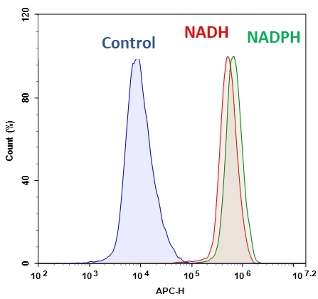
  3. Add 1 µL of JJ1902 NAD(P)H Sensor (Component A) into 0.5 mL cell suspension. Incubate at 37 oC for 30-60 minutes. Note: For a NADH/NADPH positive control treatment: Jurkat cells were incubated with 100 µM NADH or NADPH for 30 minutes in serum-free medium, and co-incubated with JJ1902 NAD(P)H Sensor working solution at 37 oC for another 30 minutes. See Figure 1 for details.

  4. Wash cells with your desired buffer such as HHBS or DPBS once. Keep cells in Assay Buffer (Component B).

  5. Monitor the fluorescence intensity at APC channel using a flow cytometer. Gate on the cells of interest, excluding debris.

Citations

View all 57 citations: Citation Explorer
Dichloroacetate radiosensitizes hypoxic breast cancer cells
Authors: de Mey, Sven and Dufait, In{\`e}s and Jiang, Heng and Corbet, Cyril and Wang, Hui and Van De Gucht, Melissa and Kerkhove, Lisa and Law, Ka Lun and Vandenplas, Hugo and Gevaert, Thierry and others,
Journal: International journal of molecular sciences (2020): 9367
Resveratrol attenuates excessive ethanol exposure induced insulin resistance in rats via improving NAD+/NADH ratio
Authors: Luo, Gang and Huang, Bingqing and Qiu, Xiang and Xiao, Lin and Wang, Ning and Gao, Qin and Yang, Wei and Hao, Liping
Journal: Molecular Nutrition & Food Research (2017)
Epigenetic regulation of Runx2 transcription and osteoblast differentiation by nicotinamide phosphoribosyltransferase
Authors: Ling, Min and Huang, Peixin and Islam, Shamima and Heruth, Daniel P and Li, Xuanan and Zhang, Li Qin and Li, Ding-You and Hu, Zhaohui and Ye, Shui Qing
Journal: Cell & Bioscience (2017): 27
MCU-dependent mitochondrial Ca2+ inhibits NAD+/SIRT3/SOD2 pathway to promote ROS production and metastasis of HCC cells
Authors: Ren, T and Zhang, H and Wang, J and Zhu, J and Jin, M and Wu, Y and Guo, X and Ji, L and Huang, Q and Yang, H and others, undefined
Journal: Oncogene (2017)
Metabolic and molecular insights into an essential role of nicotinamide phosphoribosyltransferase
Authors: Zhang, Li Q and Van Ha, undefined and el, Leon and Xiong, Min and Huang, Peixin and Heruth, Daniel P and Bi, Charlie and Gaedigk, Roger and Jiang, Xun and Li, Ding-You and Wyckoff, Gerald and others, undefined
Journal: Cell Death & Disease (2017): e2705

References

View all 1 references: Citation Explorer
Inhibition of leucine aminopeptidase 3 suppresses invasion of ovarian cancer cells through down-regulation of fascin and MMP-2/9
Authors: Wang X, Shi L, Deng Y, Qu M, Mao S, Xu L, Xu W, Fang C.
Journal: Eur J Pharmacol (2015): 116
Page updated on September 19, 2024

Ordering information

Price
Unit size
Catalog Number15296
Quantity
Add to cart

Additional ordering information

Telephone1-800-990-8053
Fax1-800-609-2943
Emailsales@aatbio.com
InternationalSee distributors
Bulk requestInquire
Custom sizeInquire
Technical SupportContact us
Purchase orderSend to sales@aatbio.com
ShippingStandard overnight for United States, inquire for international
Request quotation

Storage, safety and handling

H-phraseH303, H313, H333
Hazard symbolXN
Intended useResearch Use Only (RUO)
R-phraseR20, R21, R22
UNSPSC12352200

Platform

Flow cytometer

Excitation640 nm laser
Emission660, 20 nm filter
Instrument specification(s)APC channel

Components

Flow cytometric analysis of NADH/NADPH measurement in Jurkat cells using Cell Meter™ Intracellular NADH/NADPH Flow Cytometric Analysis Kit (Cat#15296). Cells were incubated with or without 100 µM NADH in serum-free medium for 30 minutes and then co-incubated with JJ1902 NAD(P)H sensor working solution for another 30 minutes. Fluorescence intensity was measured using ACEA NovoCyte flow cytometer in APC channel.