Products
Services
Resources
Selection Guides
About
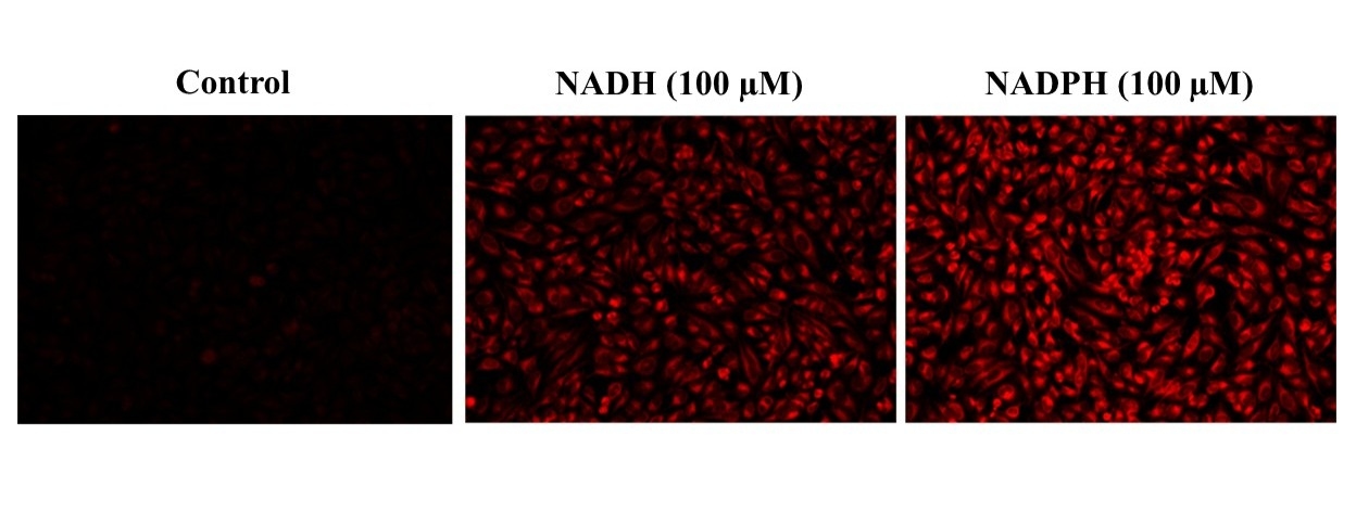
Cell Meter™ Intracellular NADH/NADPH Fluorescence Imaging Kit
Deep Red Fluorescence
The detection of intracellular dihydronicotinamide adenine dinucleotide NADH and its phosphate ester NADPH is important for disease diagnostics and drug discovery. In general, the redox couples NAD/NADH and NADP/NADPH play a critical role in energy metabolism, glycolysis, tricarboxylic acid cycle and mitochondrial respiration. The increased NAD(P)H level in cells is linked to the abnormal production of reactive oxygen species (ROS) and DNA damage. However, due to the lack of sensitive NAD(P)H probe, it has been challenging to detect intracellular NAD(P)H in biological systems. Cell Meter™ Intracellular NADH/NADPH Fluorescence Imaging Kit provides an efficient method to monitor intracellular NAD(P)H level in live cells in the far spectrum and can be combined with other applications such as GFP-expressed cells or application of MitoTracker. JJ1902 NAD(P)H sensor has been developed as an excellent fluorescent probe for detecting and imaging NADH/NADPH in cells. The probe which is fluorogenic in nature, binds NADH/NADPH to generate strong fluorescence signal with high sensitivity and specificity. JJ1902 NAD(P)H sensor can be readily loaded into live cells, and its fluorescence signal can be conveniently monitored using the filter set of Cy5®. This kit is optimized for fluorescence imaging and microplate reader applications.
| Catalog | Size | Price | Quantity |
|---|---|---|---|
| 15295 | 100 Tests | Price |

Spectral properties
| Excitation (nm) | 572 |
| Emission (nm) | 650 |
Usage and storage
| Intended use | Research Use Only (RUO) |
Instrument settings
| Fluorescence microscope | |
| Excitation | 590 nm |
| Emission | 655 nm |
| Recommended plate | Black wall/clear bottom |
| Instrument specification(s) | Cy5 filter |
| Fluorescence microplate reader | |
| Excitation | 590 nm |
| Emission | 655 nm |
| Cutoff | 610 nm |
| Recommended plate | Black wall/clear bottom |
| Instrument specification(s) | Bottom read mode |
Contact us
| Telephone | |
| Fax | |
| sales@aatbio.com | |
| International | See distributors |
| Bulk request | Inquire |
| Custom size | Inquire |
| Technical Support | Contact us |
| Request quotation | Request |
| Purchase order | Send to sales@aatbio.com |
| Shipping | Standard overnight for United States, inquire for international |
Page updated on July 17, 2026