Products
Services
Resources
Selection Guides
About
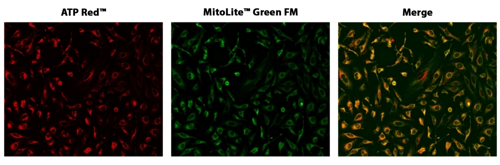
Cell Meter™ Live Cell ATP Assay Kit
Adenosine triphosphate (ATP) plays a fundamental role in cellular energetics, metabolic regulation and cellular signaling. It is referred as the "molecular unit of currency" of intracellular energy transfer to drive many biological processes and chemical synthesis in living cells. ATP also serves as a signaling molecule for cell communication and plays an important role in DNA and RNA synthesis. It is localized in mitochondria, where cellular respiration occurs. ATP levels can be used to measure cell proliferation and cell cycle dynamics. AAT Bioquest offers a variety of bioluminescence, fluorescence and colorimetric assay kits to determine ATP level in solutions. Cell Meter™ Live Cell ATP Assay Kit enables researchers to monitor ATP levels in live cells using ATP Red™, a cell-permeable red fluorescent imaging probe for detecting ATP. ATP Red™ is designed to monitor ATP concentrations in the mitochondria of living cells. The probe has minimal cross reactivity to AMP, ADP, CMP, CDP, CTP, UMP, UDP, UTP, GMP, GDP or GTP.
| Catalog | Size | Price | Quantity |
|---|---|---|---|
| 23015 | 100 Tests | Price |
Usage and storage
| Intended use | Research Use Only (RUO) |
Instrument settings
| Fluorescence microscope | |
| Excitation | Cy3/TRITC filter set |
| Emission | Cy3/TRITC filter set |
| Recommended plate | Black wall/clear bottom |
Contact us
| Telephone | |
| Fax | |
| sales@aatbio.com | |
| International | See distributors |
| Bulk request | Inquire |
| Custom size | Inquire |
| Technical Support | Contact us |
| Request quotation | Request |
| Purchase order | Send to sales@aatbio.com |
| Shipping | Standard overnight for United States, inquire for international |
Page updated on July 26, 2026