logo
AAT Bioquest

Cell Meter™ Mitochondrial Hydroxyl Radical Detection Kit *Red Fluorescence*

The detection of intracellular hydroxyl radical is of central importance to understanding proper cellular redox regulation and the impact of its dysregulation on various pathologies. The hydroxyl radical ('OH) is one of the reactive oxygen species (ROS) highly reactive with other molecules to achieve stability. In general, hydroxyl radical is considered to be a harmful by-product of oxidative metabolism, which can cause molecular damage in living system. It shows an average lifetime of 10-9 nano seconds and can react with nearly every biomolecule such as nuclear DNA, mitochondrial DNA, proteins and membrane lipids. AAT Bioquest's Cell Meter™ Mitochondrial Hydroxyl Radical Detection Kit is optimized for detecting hydroxyl radical in mitochondria. MitoROS™ OH580 is live-cell permeant probe and can rapidly and selectively target hydroxyl radical in live cells. It generates red fluorescence when it reacts with 'OH, and can be easily read. Cell Meter™ Mitochondrial Hydroxyl Radical Detection Kit provides a sensitive fluorimetric probe to detect OH' in live cells with one hour incubation. This kit can be used for fluorescence microplate readers and fluorescence microscopy applications.
Example protocol

AT A GLANCE

Protocol summary

  1. Prepare cells
  2. Incubate cells with MitoROS™ OH580 working solution at 37°C for 60 minutes
  3. Incubate cells with test compounds (to induce OH-)
  4. Monitor the fluorescence increase at Ex/Em= 540/590 nm

Important notes
Thaw all the components at room temperature before use.

PREPARATION OF STOCK SOLUTION

Unless otherwise noted, all unused stock solutions should be divided into single-use aliquots and stored at -20 °C after preparation. Avoid repeated freeze-thaw cycles.

1. MitoROS™ OH580 stock solution (500X):
Add 50 µL of DMSO (Component C) into the vial of MitoROS™ OH580 (Component A), and mix them well. Note: 25 uL of stock solution is enough for 1 plate. Note: Unused portion can be aliquoted and stored at ≤ -20ºC for more than one month if the tubes are sealed tightly and kept from light. Avoid repeated freeze-thaw cycles.

PREPARATION OF WORKING SOLUTION

Add 25 μL of 500X DMSO reconstituted MitoROS™ OH580 stock solution into 10 mL of Assay Buffer (Component B). Mix well. Note: This working solution is stable for at least 2 hours at room temperature.

For guidelines on cell sample preparation, please visit
https://www.aatbio.com/resources/guides/cell-sample-preparation.html

SAMPLE EXPERIMENTAL PROTOCOL

  1. Remove medium, and add 100 µL/well (96-well plate) or 25 µL/well (384-well plate) of MitoROS™ OH580 working solution into the cell plate. Incubate cells at 37°C for 60 minutes.

  2. To induce hydroxyl radical, treat cells with test compounds in your desired buffer (such as PBS or HHBS) at 37°C for a desired period of time, protected from light. Note: We treated HeLa cells with Fenton reaction (10 µM CuCl2 and 100 µM H2O2) at 37°C for 1 hour to induce exogenous hydroxyl radical. See Figure 1 for details. We treated RAW 264.7 cells with PMA (phorbol 12-myristate 13-acetate) in growth medium at 37°C for 4 hours to stimulate endogenous hydroxyl radical.

  3. Wash cells 2 - 3 times with HHBS or DPBS, and add 100 µL Assay Buffer (Component B) to each well.

  4. Monitor the fluorescence signal in cells using fluorescence microscope with a TRITC filter set, or measure fluorescence increase using fluorescence microplate reader at Ex/Em = 540/590 nm (cut off = 570 nm) with bottom read mode.

Spectrum

Citations

View all 5 citations: Citation Explorer
Tetrandrine Targeting SIRT5 Exerts Anti-melanoma Properties via Inducing ROS, ER Stress, and Blocked Autophagy
Authors: Ji, Yacong and Li, Chongyang and Wan, Sicheng and Dong, Zhen and Liu, Chaolong and Guo, Leiyang and Shi, Shaomin and Ci, Mingxin and Xu, Minghao and Li, Qian and others,
Journal: Journal of Pharmaceutical Analysis (2024): 101036
Hydrogen peroxide-induced oxidative stress promotes expression of CXCL15/Lungkine mRNA in a MEK/ERK-dependent manner in fibroblast-like synoviocytes derived from mouse temporomandibular joint
Authors: Asanuma, Kanna and Yokota, Seiji and Chosa, Naoyuki and Kamo, Masaharu and Ibi, Miho and Mayama, Hisayo and Iri{\'e}, Tarou and Satoh, Kazuro and Ishisaki, Akira
Journal: Journal of Oral Biosciences (2022)
A New Drug-Free Cancer Therapy Using Ultraviolet Pulsed Irradiation. PDT (PhotoDynamic Therapy) to PPT (Pulsed Photon Therapy)
Authors: Itoh, Johbu and Itoh, Yoshiko
Journal: Frontiers in Bioscience-Scholar (2022): 27
Tetrahydrofolate alleviates the inhibitory effect of oxidative stress on neural stem cell proliferation through PTEN/Akt/mTOR pathway
Authors: Zhang, Xuyang and Liu, Zhi and Yang, Wenqin and Zhao, Fengchun and Zhang, Chao and Feng, Hui and Zhou, Tengyuan and Zhong, Jun and Zou, Yongjie and Feng, Hua and others,
Journal: Oxidative Medicine and Cellular Longevity (2022)
Homogeneously catalytic oxidation of phenanthrene by the reaction of extracellular secretions of pyocyanin and Nicotinamide Adenine Dinucleotide
Authors: Nie, Hongyun and Nie, Maiqian and Diwu, Zhenjun and Wang, Lei and Qiao, Qi and Zhang, Bo and Yang, Xuefu
Journal: Environmental Research (2020): 110159

References

View all 47 references: Citation Explorer
Oxyl and hydroxyl radical transfer in mitochondrial amidoxime reducing component-catalyzed nitrite reduction
Authors: Yang J, Giles LJ, Ruppelt C, Mendel RR, Bittner F, Kirk ML.
Journal: J Am Chem Soc (2015): 5276
Arbutin, an intracellular hydroxyl radical scavenger, protects radiation-induced apoptosis in human lymphoma U937 cells
Authors: Wu LH, Li P, Zhao QL, Piao JL, Jiao YF, Kadowaki M, Kondo T.
Journal: Apoptosis (2014): 1654
Chloroplast-located BjFer1 together with anti-oxidative genes alleviate hydrogen peroxide and hydroxyl radical injury in cytoplasmic male-sterile Brassica juncea
Authors: Yang J, Liu S, Yang X, Zhang M.
Journal: Mol Biol Rep (2012): 4169
Hydroxyl radical (.OH) played a pivotal role in oridonin-induced apoptosis and autophagy in human epidermoid carcinoma A431 cells
Authors: Yu Y, Fan SM, Song JK, Tashiro S, Onodera S, Ikejima T.
Journal: Biol Pharm Bull (2012): 2148
Excess no predisposes mitochondrial succinate-cytochrome c reductase to produce hydroxyl radical
Authors: Chen J, Chen CL, Alevriadou BR, Zweier JL, Chen YR.
Journal: Biochim Biophys Acta (2011): 491
Page updated on September 8, 2024

Ordering information

Price
Unit size
Catalog Number16055
Quantity
Add to cart

Additional ordering information

Telephone1-800-990-8053
Fax1-800-609-2943
Emailsales@aatbio.com
InternationalSee distributors
Bulk requestInquire
Custom sizeInquire
Technical SupportContact us
Purchase orderSend to sales@aatbio.com
ShippingStandard overnight for United States, inquire for international
Request quotation

Spectral properties

Excitation (nm)

576

Emission (nm)

598

Storage, safety and handling

H-phraseH303, H313, H333
Hazard symbolXN
Intended useResearch Use Only (RUO)
R-phraseR20, R21, R22
UNSPSC12352200

Platform

Fluorescence microscope

ExcitationCy3, TRITC filter set
EmissionCy3, TRITC filter set
Recommended plateBlack wall, clear bottom

Fluorescence microplate reader

Excitation540 nm
Emission590 nm
Cutoff570 nm
Recommended plateBlack wall, clear bottom
Instrument specification(s)Bottom read mode

Components

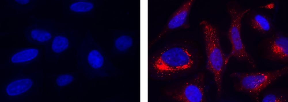
Fluorescence images of hydroxyl radical measurement in HeLa cells using MitoROS&trade; OH580 (Cat#16055). HeLa cells were incubated with MitoROS&trade; OH580 working solution at 37 &deg;C for 1 hour, then washed once with HHBS. Fenton Reaction: Cells were then treated with 10 &micro;M CuCl2 and 100 &micro;M H<sub>2</sub>O<sub>2</sub> in 1X HBSS buffer at 37 &deg;C for 1 hour. Control: HeLa cells were kept in 1X HBSS buffer without treatment. After washing 3 times with HHBS, HeLa cells were measured using a fluorescence microscope with a TRITC filter set (Red). Cell nuclei were stained with Hoechst 33342 (Cat#17530, Blue).
Detection of intracellular hydroxyl radical in RAW 264.7 cells using MitoROS&trade;OH580 (Cat#16055). Cells were incubated with MitoROS&trade; OH580 working solution at 37 &ordm;C for 1 hour, then washed once with HHBS. Cells were then incubated without or with PMA (phorbol 12-myristate 13-acetate, 10 to 500 ng/mL) in growth medium at 37&ordm;C for 4 hours. After washing 3 times with HHBS, HeLa cells were measured using a fluorescence microscope with a TRITC filter set.
Antioxidant NAC partially and significantly reverses the H2O2-mediated increase in phosphorylation of ERK1/2 in FLS1 cells. (A) Cells were cultured (Aa) without or (Ab) with H2O2 (500 μM) for 24 h, and then OH∙ production in the cells was fluorescently visualized. Detection of hydroxyl radicals in FLS1 cells was performed using a Cell Meter™ mitochondrial hydroxyl radical (OH∙) detection kit under a fluorescence microscope. The nuclei were counter-stained with Hoechst 33342 (blue). Scale bar, 50 μm. Source: <b>Hydrogen peroxide-induced oxidative stress promotes expression of CXCL15/Lungkine mRNA in a MEK/ERK-dependent manner in fibroblast-like synoviocytes derived from mouse temporomandibular joint</b> by Asanuma, Kanna et.al., <em>Journal of Oral Biosciences</em>, March 2023
Cellular mitochondrial ROS-OH580 activity under 24 h pulsed UV irradiation in MCF-7 and Cos7 cells. (A) The signal intensities of PI and ROS-OH580 in control MCF-7 cells were weakly distributed in the cytoplasm and nucleus. Bar = 5 μm. Cell atrophy and cytoplasmic lysis were observed in irradiated MCF-7 cells. Most cells were PI-positive (cell death), and the ROS-OH580 (•OH) signal was strongly detected in the nuclear enrichment. Bar = 5μm. (B) PI and ROS-OH580 (•OH) signal intensities in Cos7 cells were weakly distributed in the cytoplasm of control and irradiated Cos7 cells. Bar = 5μm. Source: <b>A New Drug-Free Cancer Therapy Using Ultraviolet Pulsed Irradiation. PDT (PhotoDynamic Therapy) to PPT (Pulsed Photon Therapy).</b> by Johbu Itoh, Yoshiko Itoh. <em>Front. Biosci. (Schol Ed)</em>, Sept. 2022.