Cell Meter™ Multiplexing Live, Apoptotic and Necrotic Cell Detection Kit III *Triple Fluorescence Colors*
Price | |
Catalog Number | |
Unit Size | |
Quantity |
Telephone | 1-800-990-8053 |
Fax | 1-800-609-2943 |
sales@aatbio.com | |
Quotation | Request |
International | See distributors |
Shipping | Standard overnight for United States, inquire for international |
H-phrase | H303, H313, H333 |
Hazard symbol | XN |
Intended use | Research Use Only (RUO) |
R-phrase | R20, R21, R22 |
UNSPSC | 12352200 |
Overview | ![]() ![]() |
Platform
Fluorescence microscope
Excitation | 494 nm (apoptosis) / 350 nm (necrosis) / 613 nm (live) |
Emission | 520 nm (apoptosis) / 461 nm (necrosis) / 631 nm (live) |
Recommended plate | Black wall/clear bottom |
Instrument specification(s) | FITC filter (apoptosis) / DAPI filter (necrosis) / Cy5 filter (live) |
Components
Example protocol
AT A GLANCE
Protocol summary
- Prepare cells with test compounds
- Add triple fluorescence working solution (100 µL/sample)
- Incubate at room temperature or 37oC for 30 - 60 minutes
- Analyze with a fluorescence microscope at FITC channel (apoptosis), DAPI channel (necrosis) or Texas Red/Cy5 channel (healthy cells)
Important notes
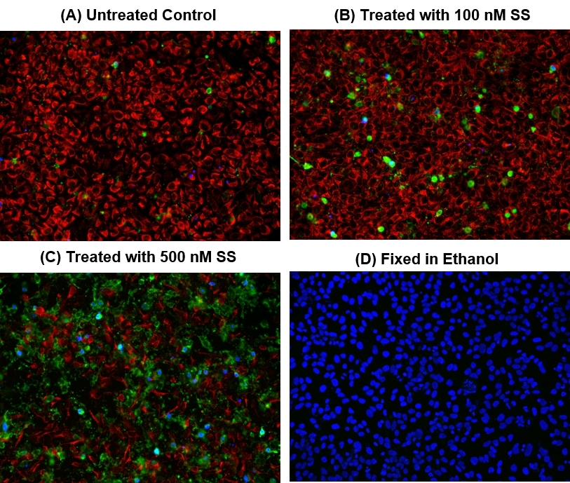
We treated HeLa cells with staurosporine (SS) for 4 hours at 37oC to induce cell apoptosis. See Figure 1 for details.
Thaw all components to room temperature before beginning protocol.
PREPARATION OF WORKING SOLUTION
Add 10 µL of 100X Annexin V-iFluor™ 488 conjugate (Component A), 5 µL of 200X Nuclear Blue™ DCS1 (Component C) and 5 µL of 200X Cellbrite™ Red (Component D) to 1 mL of Assay Buffer (Component B). The triple fluorescence assay solution is stable for at least 1 hour at room temperature. Note: As the optimal staining conditions may vary depending on different cell types, it’s recommended to determine the appropriate concentration of Component A, C and D individually.
For guidelines on cell sample preparation, please visit
https://www.aatbio.com/resources/guides/cell-sample-preparation.html
SAMPLE EXPERIMENTAL PROTOCOL
- Remove cell culture medium and test compounds after treatment.
- Add 100 µL/well (96-well plate) or 50 µL/well (384-well plate) of triple fluorescence assay solution. Incubate at 37oC for 30 to 60 minutes, protected from light.
- Wash cells with HBSS, PBS or buffer of your choice twice.
- Analyze the apoptotic cells with Annexin V-iFluor™ 488 conjugate under fluorescence microscope with a FITC filter. The green staining (Ex/Em = 494/520 nm) on the plasma membrane indicates the Annexin V-iFluor™ 488 conjugate binding to PS on cell surface. Monitor the fluorescence intensity with a DAPI filter (Ex/Em = 350/461 nm) for necrosis, Texas Red or Cy5 filter (Ex/Em = 613/631 nm) for live cells using a fluorescence microscope (See Figure 1 for details).
Images

Citations
Authors: Takada, Ichiro and Hidano, Shinya and Takahashi, Sayuri and Yanaka, Kaori and Ogawa, Hidesato and Tsuchiya, Megumi and Yokoyama, Atsushi and Sato, Shingo and Ochi, Hiroki and Nakagawa, Tohru and others,
Journal: Journal of Biological Chemistry (2022)
Authors: Nanashima, Naoki and Horie, Kayo and Chiba, Mitsuru and Nakano, Manabu and Maeda, Hayato and Nakamura, Toshiya
Journal: Molecular Medicine Reports (2017): 6134--6141
Authors: Riaz, Muhammad Assad and Stammler, Angelika and Borgers, Mareike and Konrad, Lutz
Journal: American Journal of Translational Research (2017): 1266
Authors: Coleman, Jack and Liu, Rui and Wang, Kathy and Kumar, Arun
Journal: Apoptosis Methods in Toxicology (2016): 77--92
References
Authors: Zhang M, Zhou L.
Journal: Zhonghua Er Bi Yan Hou Tou Jing Wai Ke Za Zhi (2009): 565
Authors: Ahmed S, Silverman MD, Marotte H, Kwan K, Matuszczak N, Koch AE.
Journal: Arthritis Rheum (2009): 1282
Authors: Hong S, Kim CY, Lee JE, Seong GJ.
Journal: Life Sci (2009): 28
Authors: Selzner M, Selzner N, Chen L, Borozan I, Sun J, Xue-Zhong M, Zhang J, McGilvray ID.
Journal: Liver Transpl (2009): 1594
Authors: Hong JY, Lebofsky M, Farhood A, Jaeschke H.
Journal: Am J Physiol Gastrointest Liver Physiol (2009): G572
Authors: Ling S, Luo R, Dai A, Guo Z, Guo R, Komesaroff PA.
Journal: Phytomedicine (2009): 56
Authors: Shi H, Guan SH.
Journal: Liver Int (2009): 349
Authors: Meusch U, Rossol M, Baerwald C, Hauschildt S, Wagner U.
Journal: Arthritis Rheum (2009): 2612
Authors: Sisto M, D'Amore M, Caprio S, Mitolo V, Scagliusi P, Lisi S.
Journal: Ann N Y Acad Sci (2009): 407
Authors: Elders RC, Baines SJ, Catchpole B.
Journal: Vet Immunol Immunopathol (2009): 11
Application notes
FAQ
Do you offer any fluorimetric assays that measure caspase activation/activity in live cells using a flow cytometer?
Does pH and staining temperature affect Annexin V-Phosphatidylserine binding?
Does propidium iodide stain apoptotic cells?
How can I tell if my cell sample is dying?
AssayWise
Nucleic Acid Detection, Quantification and Imaging
A practical guide for use of PE and APC in flow cytometry
Fluorescent Phalloidin: A Practical Stain for Visualizing Actin Filaments
Power Styramide™ Signal Amplification a Superior Alternative to Tyramide Signal Amplification