logo
AAT Bioquest

Cell Meter™ Multiplexing Live, Apoptotic and Necrotic Cell Detection Kit III *Triple Fluorescence Colors*

Our Cell Meter™ assay kits are a set of tools for monitoring cell viability. There are a variety of parameters that can be used. This particular kit is designed to simultaneously monitor apoptotic, necrotic and healthy cells. Apoptosis is an active, programmed process of autonomous cellular dismantling that avoids eliciting inflammation. In apoptosis, phosphatidylserine (PS) is transferred to the outer leaflet of the plasma membrane. As a universal indicator of the initial/intermediate stages of cell apoptosis, the appearance of phosphatidylserine on the cell surface can be detected before morphological changes are observed. The PS sensor Annexin V-iFluor® 488 conjugate has green fluorescence upon binding to membrane PS. Necrosis has been characterized as passive, accidental cell death resulting from environmental perturbations with uncontrolled release of inflammatory cellular contents. Loss of plasma membrane integrity, as demonstrated by the ability of a membrane-impermeable Nuclear Blue™ DCS1 (Ex/Em = 350/461 nm) to label the nucleus, represents a straightforward approach to demonstrate late stage of apoptosis and necrosis. In addition, this kit also provides a live cell labeling dye, Cellbrite™ Red (Ex/Em = 613/631 nm), for labeling non-apoptotic healthy cells. This kit is optimized to simultaneously detect cell apoptosis (green), necrosis (blue and/or green) and healthy cells (red) with a fluorescence microscope.
Example protocol

AT A GLANCE

Protocol summary

  1. Prepare cells with test compounds
  2. Add triple fluorescence working solution (100 µL/sample)
  3. Incubate at room temperature or 37oC for 30 - 60 minutes
  4. Analyze with a fluorescence microscope at FITC channel (apoptosis), DAPI channel (necrosis) or Texas Red/Cy5 channel (healthy cells)

Important notes
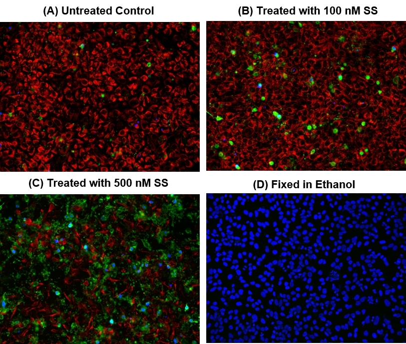
We treated HeLa cells with staurosporine (SS) for 4 hours at 37oC to induce cell apoptosis. See Figure 1 for details.
Thaw all components to room temperature before beginning protocol.

PREPARATION OF WORKING SOLUTION

Add 10 µL of 100X Annexin V-iFluor™ 488 conjugate (Component A), 5 µL of 200X Nuclear Blue™ DCS1 (Component C) and 5 µL of 200X Cellbrite™ Red (Component D) to 1 mL of Assay Buffer (Component B). The triple fluorescence assay solution is stable for at least 1 hour at room temperature. Note: As the optimal staining conditions may vary depending on different cell types, it’s recommended to determine the appropriate concentration of Component A, C and D individually.

For guidelines on cell sample preparation, please visit
https://www.aatbio.com/resources/guides/cell-sample-preparation.html

SAMPLE EXPERIMENTAL PROTOCOL

  1. Remove cell culture medium and test compounds after treatment.

  2. Add 100 µL/well (96-well plate) or 50 µL/well (384-well plate) of triple fluorescence assay solution. Incubate at 37oC for 30 to 60 minutes, protected from light.

  3. Wash cells with HBSS, PBS or buffer of your choice twice.

  4. Analyze the apoptotic cells with Annexin V-iFluor™ 488 conjugate under fluorescence microscope with a FITC filter. The green staining (Ex/Em = 494/520 nm) on the plasma membrane indicates the Annexin V-iFluor™ 488 conjugate binding to PS on cell surface. Monitor the fluorescence intensity with a DAPI filter (Ex/Em = 350/461 nm) for necrosis, Texas Red or Cy5 filter (Ex/Em = 613/631 nm) for live cells using a fluorescence microscope (See Figure 1 for details).

Citations

View all 4 citations: Citation Explorer
Transcriptional coregulator Ess2 controls survival of post-thymic CD4+ T cells through the Myc and IL-7 signaling pathways
Authors: Takada, Ichiro and Hidano, Shinya and Takahashi, Sayuri and Yanaka, Kaori and Ogawa, Hidesato and Tsuchiya, Megumi and Yokoyama, Atsushi and Sato, Shingo and Ochi, Hiroki and Nakagawa, Tohru and others,
Journal: Journal of Biological Chemistry (2022)
Anthocyanin-rich blackcurrant extract inhibits proliferation of the MCF10A healthy human breast epithelial cell line through induction of G0/G1 arrest and apoptosis
Authors: Nanashima, Naoki and Horie, Kayo and Chiba, Mitsuru and Nakano, Manabu and Maeda, Hayato and Nakamura, Toshiya
Journal: Molecular Medicine Reports (2017): 6134--6141
Clusterin signals via ApoER2/VLDLR and induces meiosis of male germ cells
Authors: Riaz, Muhammad Assad and Stammler, Angelika and Borgers, Mareike and Konrad, Lutz
Journal: American Journal of Translational Research (2017): 1266
Detecting Apoptosis, Autophagy, and Necrosis
Authors: Coleman, Jack and Liu, Rui and Wang, Kathy and Kumar, Arun
Journal: Apoptosis Methods in Toxicology (2016): 77--92

References

View all 71 references: Citation Explorer
Tumor necrosis factor inhibitors block apoptosis of human epithelial cells of the salivary glands
Authors: Sisto M, D'Amore M, Caprio S, Mitolo V, Scagliusi P, Lisi S.
Journal: Ann N Y Acad Sci (2009): 407
PVP-coated silver nanoparticles and silver ions induce reactive oxygen species, apoptosis and necrosis in THP-1 monocytes
Authors: Foldbjerg R, Olesen P, Hougaard M, Dang DA, Hoffmann HJ, Autrup H.
Journal: Toxicol Lett (2009): 156
Concurrent induction of necrosis, apoptosis, and autophagy in ischemic preconditioned human livers formerly treated by chemotherapy
Authors: Domart MC, Esposti DD, Sebagh M, Olaya N, Harper F, Pierron G, Franc B, Tanabe KK, Debuire B, Azoulay D, Brenner C, Lemoine A.
Journal: J Hepatol (2009): 881
Rapid simultaneous determination of apoptosis, necrosis, and viability in sulfur mustard exposed HaCaT cell cultures
Authors: Heinrich A, Balszuweit F, Thiermann H, Kehe K.
Journal: Toxicol Lett (2009): 260
RAS mutations affect tumor necrosis factor-induced apoptosis in colon carcinoma cells via ERK-modulatory negative and positive feedback circuits along with non-ERK pathway effects
Authors: Kreeger PK, M and hana R, Alford SK, Haigis KM, Lauffenburger DA.
Journal: Cancer Res (2009): 8191
Page updated on September 8, 2024

Ordering information

Price
Unit size
Catalog Number22846
Quantity
Add to cart

Additional ordering information

Telephone1-800-990-8053
Fax1-800-609-2943
Emailsales@aatbio.com
InternationalSee distributors
Bulk requestInquire
Custom sizeInquire
Technical SupportContact us
Purchase orderSend to sales@aatbio.com
ShippingStandard overnight for United States, inquire for international
Request quotation

Storage, safety and handling

H-phraseH303, H313, H333
Hazard symbolXN
Intended useResearch Use Only (RUO)
R-phraseR20, R21, R22
UNSPSC12352200

Platform

Fluorescence microscope

Excitation494 nm (apoptosis) , 350 nm (necrosis) , 613 nm (live)
Emission520 nm (apoptosis) , 461 nm (necrosis) , 631 nm (live)
Recommended plateBlack wall, clear bottom
Instrument specification(s)FITC filter (apoptosis) , DAPI filter (necrosis) , Cy5 filter (live)

Components

Fluorescence images of HeLa cells labeled with Cell Meter&trade; Multiplexing Live, Apoptotic and Necrotic Detection Kit *Triple Fluorescence* (Cat#22846). HeLa cells at 100,000 cells/well/100 &micro;L were seeded overnight in a 96-well black wall/clear bottom plate. Cells were treated with 0-500 nM staurosporine (SS) at&nbsp;37<sup>o</sup>C for 4 hours (A-C), or fixed in ethanol (D), then incubated with triple fluorescence assay solution for 1 hour. The fluorescence signal was measured using a fluorescence microscope with a Cy5 filter for healthy cells (Red), FITC filter for apoptotic (Green) and DAPI filter for necrotic cells (Blue), respectively.