Products
Services
Resources
Selection Guides
About
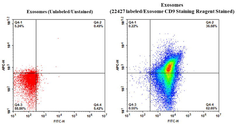
Cell Navigator® Exosome Fluorescence Staining Kit
Orange Fluorescence
Cell Navigator® Exosome Fluorescence Staining Kit (Orange Fluorescence) is a reliable tool for labeling extracellular vesicles with Exsomight™ Orange dye for clear, low-background detection in fNTA, flow cytometry, and fluorescence microscopy.
- Minimized aggregation and background: Enhances solubility to reduce dye clustering and non-specific fluorescence
- Efficient EV membrane integration: Provides uniform labeling with high signal fidelity and minimal off-target staining
- Applications: Ideal for EV population analysis, cellular uptake studies, and high-resolution imaging
- Comparable alternative: Offers a comparable option to PKH26-based EV labeling kits from Sigma-Aldrich with reduced aggregation and improved detection accuracy
| Catalog | Size | Price | Quantity |
|---|---|---|---|
| 22427 | 100 Tests | Price |
Usage and storage
| Intended use | Research Use Only (RUO) |
Contact us
| Telephone | |
| Fax | |
| sales@aatbio.com | |
| International | See distributors |
| Bulk request | Inquire |
| Custom size | Inquire |
| Technical Support | Contact us |
| Request quotation | Request |
| Purchase order | Send to sales@aatbio.com |
| Shipping | Standard overnight for United States, inquire for international |
Page updated on July 17, 2026