logo
AAT Bioquest

Cell Navigator® F-Actin Labeling Kit *Orange Fluorescence*

Our Cell Navigator® fluorescence imaging kits are a set of fluorescence imaging tools for labeling sub-cellular organelles such as membranes, lysosomes, mitochondria and nuclei etc. The selective labeling of live cell compartments provides a powerful method for studying cellular events in a spatial and temporal context. This particular kit is designed to label F-actins of fixed cells in red fluorescence. The kit uses a n orange fluorescent phalloidin conjugate that is selectively bound to F-actins. This red fluorescent phalloidin conjugate is a high-affinity probe for F-actins. Used at nanomolar concentrations, phallotoxins are convenient probes for labeling, identifying and quantitating F-actins in formaldehyde-fixed and permeabilized tissue sections, cell cultures or cell-free experiments. The labeling protocol is robust, requiring minimal hands-on time. The kit provides all the essential components with an optimized staining protocol.
Example protocol

AT A GLANCE

Protocol summary

  1. Prepare samples (microplate wells)
  2. Remove the liquid from the plate
  3. Add 100 µL/well of iFluor™ 546-Phalloidin working solution
  4. Incubate the cells at room temperature for 15 to 60 minutes
  5. Wash the cells
  6. Examine the specimen under fluorescence microscope at Ex/Em = 550/575 nm (TRITC filter set)

Important notes
Thaw all the components at room temperature before starting the experiment.

PREPARATION OF WORKING SOLUTION

Add 10 μL of iFluor™ 546-Phalloidin (Component A) to 10 mL of Labeling Buffer (Component B) to make 1X iFluor™ 546-Phalloidin working solution. Protect from light. Note: Different cell types might be stained differently. The concentration of iFluor™ 546-Phalloidin working solution should be prepared accordingly.

For guidelines on cell sample preparation, please visit
https://www.aatbio.com/resources/guides/cell-sample-preparation.html

SAMPLE EXPERIMENTAL PROTOCOL

  1. Perform formaldehyde fixation. Incubate the cells with 3.0% – 4.0% formaldehyde in PBS at room temperature for 10 – 30 minutes. Note: Avoid any methanol containing fixatives since methanol can disrupt actin during the fixation process. The preferred fixative is methanol-free formaldehyde.

  2. Rinse the fixed cells 2 – 3 times in PBS.

  3. Optional: Add 0.1% Triton X-100 in PBS into fixed cells for 3 to 5 minutes to increase permeability. Rinse the cells 2 – 3 times in PBS.

  4. Add 100 µL/well (96-well plate) of iFluor™ 546-Phalloidin working solution into the fixed cells.

  5. Incubate the cells at room temperature for 15 to 60 minutes.

  6. Rinse cells gently with PBS 2 to 3 times to remove excess dye before plate sealing.

  7. Image cells using a fluorescence microscope with TRITC filter set (Ex/Em = 550/575 nm).

Spectrum

Product family

NameExcitation (nm)Emission (nm)Extinction coefficient (cm -1 M -1)Quantum yieldCorrection Factor (260 nm)Correction Factor (280 nm)
Cell Navigator® F-Actin Labeling Kit *Blue Fluorescence*3454502000010.9510.830.23
Cell Navigator® F-Actin Labeling Kit *Green Fluorescence*4915167500010.910.210.11
Cell Navigator® F-Actin Labeling Kit *Red Fluorescence*58760320000010.5310.050.04

Citations

View all 26 citations: Citation Explorer
Evolution of phase, morphology, physicochemical properties, and biological properties of HA ceramic with the increase of crystallinity
Authors: Zhang, Luhui and Liang, Xinzhi and Chen, Ji and Kang, Zhengyang and Ye, Jiandong and Xie, Denghui
Journal: Ceramics International (2024)
Temperature-Controlled Screening of Catechol Groups in Poly (N-Isopropylacrylamide-co-Dopamine Methacrylamide) for Cell Detachment
Authors: Yang, Liuxin and Ren, Pengfei and Wei, Dandan and Liang, Min and Xu, Li and Tao, Yinghua and Jiao, Guanhua and Zhang, Tianzhu and Zhang, Qianli
Journal: ACS Applied Polymer Materials (2024)
Reproduction of Entomopathogenic Nematodes for Use in Pest Control
Authors: Abd-Elgawad, Mahfouz MM
Journal: (2024): 351--382
Microfluidic Chip-Based Modeling of Three-Dimensional Intestine--Vessel--Liver Interactions in Fluorotelomer Alcohol Biotransformation
Authors: Xu, Ning and Lin, Haifeng and Lin, Jin-Ming and Cheng, Jie and Wang, Peilong and Lin, Ling
Journal: Analytical Chemistry (2023)

References

View all 42 references: Citation Explorer
Velocity distributions of single F-actin trajectories from a fluorescence image series using trajectory reconstruction and optical flow mapping
Authors: von Wegner F, Ober T, Weber C, Schurmann S, Winter R, Friedrich O, Fink RH, Vogel M.
Journal: J Biomed Opt (2008): 54018
Visualization of F-actin and G-actin equilibrium using fluorescence resonance energy transfer (FRET) in cultured cells and neurons in slices
Authors: Okamoto K, Hayashi Y.
Journal: Nat Protoc (2006): 911
The effect of F-actin on the relay helix position of myosin II, as revealed by tryptophan fluorescence, and its implications for mechanochemical coupling
Authors: Conibear PB, Malnasi-Csizmadia A, Bagshaw CR.
Journal: Biochemistry (2004): 15404
Analysis of models of F-actin using fluorescence resonance energy transfer spectroscopy
Authors: Moens PD, dos Remedios CG.
Journal: Results Probl Cell Differ (2001): 59
Fluorescence studies of the carboxyl-terminal domain of smooth muscle calponin effects of F-actin and salts
Authors: Bartegi A, Roustan C, Kassab R, Fattoum A.
Journal: Eur J Biochem (1999): 335
Page updated on September 19, 2024

Ordering information

Price
Unit size
Catalog Number22663
Quantity
Add to cart

Additional ordering information

Telephone1-800-990-8053
Fax1-800-609-2943
Emailsales@aatbio.com
InternationalSee distributors
Bulk requestInquire
Custom sizeInquire
Technical SupportContact us
Purchase orderSend to sales@aatbio.com
ShippingStandard overnight for United States, inquire for international
Request quotation

Spectral properties

Correction Factor (260 nm)

0.25

Correction Factor (280 nm)

0.15

Extinction coefficient (cm -1 M -1)

1000001

Excitation (nm)

541

Emission (nm)

557

Quantum yield

0.671

Storage, safety and handling

H-phraseH303, H313, H333
Hazard symbolXN
Intended useResearch Use Only (RUO)
R-phraseR20, R21, R22
UNSPSC12352200

Platform

Fluorescence microscope

ExcitationTRITC filter
EmissionTRITC filter
Recommended plateBlack wall, clear bottom

Components

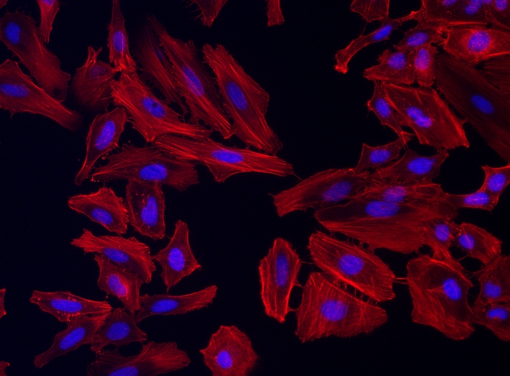
Fluorescence image of HeLa cells fixed with 4% formaldehyde then stained with Cell Navigator® F-Actin Labeling Kit *Orange Fluorescence* in a Costar black 96-well plate. Cells were labeled with iFluor® 546-Phalloidin (Cat#22663, Red) and nuclei stain DAPI (Cat#17507, Blue), respectively.  
Fluorescence image of HeLa cells fixed with 4% formaldehyde then stained with Cell Navigator® F-Actin Labeling Kit *Orange Fluorescence* in a Costar black 96-well plate. Cells were labeled with iFluor® 546-Phalloidin (Cat#22663, Red) and nuclei stain DAPI (Cat#17507, Blue), respectively.