Products
Services
Resources
Selection Guides
About
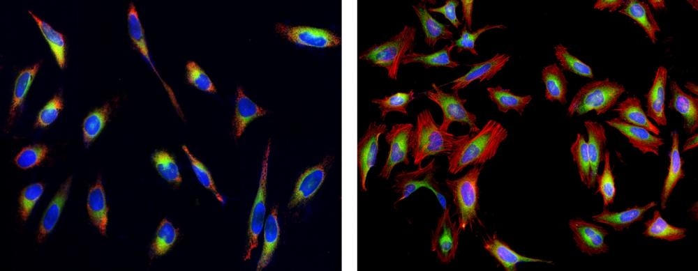
Cell Navigator® Live Cell Endoplasmic Reticulum (ER) Staining Kit
Green Fluorescence
The endoplasmic reticulum (ER) is a type of organelle in the cells of eukaryotic organisms that forms an interconnected network of flattened, membrane-enclosed sacs or tube-like structures known as cisternae. The membranes of the ER are continuous with the outer nuclear membrane. ER occurs in most types of eukaryotic cells, but is absent from red blood cells and spermatozoa. This Cell Navigator® Live Cell Endoplasmic Reticulum (ER) Staining Kit uses our ER Tracer™ Green as an ER marker. ER Tracer™ Green stain is a cell-permeant fluorescent dye that is highly selective for ER. This stain consists of a green fluorescent dye and ER binder that selectively bind to ER in most of cell types. For some cells, ER Tracer™ Green may not selectively bind to ER. ER Tracer™ Green has spectral properties essentially identical to FITC, making this kit convenient with the FITC filter set. ER Tracer™ is good replacement for ER Tracker (trademark ThermoFisher).
| Catalog | Size | Price | Quantity |
|---|---|---|---|
| 22635 | 100 Tests | Price |

Spectral properties
| Excitation (nm) | 503 |
| Emission (nm) | 511 |
Storage, safety and handling
| H-phrase | H303, H313, H333 |
| Hazard symbol | XN |
| Intended use | Research Use Only (RUO) |
| R-phrase | R20, R21, R22 |
| UNSPSC | 12352200 |
Instrument settings
| Fluorescence microscope | |
| Excitation | 490 nm |
| Emission | 520 nm |
| Recommended plate | Black wall/clear bottom |
| Instrument specification(s) | FITC filter |
Contact us
| Telephone | |
| Fax | |
| sales@aatbio.com | |
| International | See distributors |
| Bulk request | Inquire |
| Custom size | Inquire |
| Technical Support | Contact us |
| Request quotation | Request |
| Purchase order | Send to sales@aatbio.com |
| Shipping | Standard overnight for United States, inquire for international |
Page updated on June 22, 2026