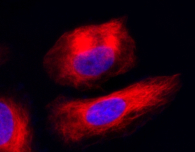
logo
AAT Bioquest

Cell Navigator® Live Cell Tubulin Staining Kit

Imaging of tubulins in live HeLa cells. HeLa cells were co-labelled with Tubulite<sup>TM</sup> Red and DAPI (Cat# 17507) for 60 minutes in a 37 <sup>o</sup>C, 5% CO<sub>2</sub> incubator. The fluorescence image was taken with a fluorescence microscope (Cy5 filter set).
Ordering information
Price
Catalog Number
Unit Size
Quantity
Add to cart
Additional ordering information
Telephone1-800-990-8053
Fax1-800-609-2943
Emailsales@aatbio.com
InternationalSee distributors
Bulk requestInquire
Custom sizeInquire
ShippingStandard overnight for United States, inquire for international
Request quotation
Storage, safety and handling
Certificate of OriginDownload PDF
Intended useResearch Use Only (RUO)
Related products
Cell Navigator® Live Cell RNA Imaging Kit *Green Fluorescence*
Cell Navigator® Live Cell Endoplasmic Reticulum (ER) Staining Kit *Green Fluorescence*
Cell Navigator® Live Cell Endoplasmic Reticulum (ER) Staining Kit *Red Fluorescence*
Cell Navigator® Lysosome Staining Kit *Green Fluorescence with 405 nm Excitation*
Cell Navigator® Lysosome Staining Kit *NIR Fluorescence*
Cell Navigator® Lysosome Staining Kit *Blue Fluorescence*
Cell Navigator® Lysosome Staining Kit *Green Fluorescence*
Cell Navigator® Lysosome Staining Kit *Orange Fluorescence*
Cell Navigator® Lysosome Staining Kit *Red Fluorescence*
Cell Navigator® Lysosome Staining Kit *Deep Red Fluorescence*
Cell Navigator® F-Actin Labeling Kit *Blue Fluorescence*
Cell Navigator® F-Actin Labeling Kit *Green Fluorescence*
Cell Navigator® F-Actin Labeling Kit *Orange Fluorescence*
Cell Navigator® F-Actin Labeling Kit *Red Fluorescence*
Cell Navigator® Mitochondrion Staining Kit *Blue Fluorescence*
Cell Navigator® Mitochondrion Staining Kit *Green Fluorescence*
Cell Navigator® Mitochondrion Staining Kit *Orange Fluorescence*
Cell Navigator® Mitochondrion Staining Kit *Red Fluorescence*
Cell Navigator® Mitochondrion Staining Kit *Deep Red Fluorescence*
Cell Navigator® Mitochondrion Staining Kit *Orange Fluorescence with 405 nm Excitation*
Cell Navigator® Cell Plasma Membrane Staining Kit *Orange Fluorescence*
Cell Navigator® Cell Plasma Membrane Staining Kit *Red Fluorescence*
Cell Navigator® Mitochondrion Staining Kit *NIR Fluorescence*
Cell Navigator® Fluorimetric Lipid Droplet Assay Kit *Green Fluorescence*
Cell Navigator® Fluorimetric Lipid Droplet Assay Kit *Red Fluorescence*
Cell Navigator® NBD Ceramide Golgi Staining Kit *Green Fluorescence*
Cell Navigator® Cell Plasma Membrane Staining Kit *Green Fluorescence*
Cell Navigator® Live Cell Endoplasmic Reticulum (ER) Staining Kit *Blue Fluorescence*
Cell Navigator® TMR Ceramide Golgi Staining Kit *Red Fluorescence*
Cell Navigator® CDy6 Mitosis Imaging Kit
Cell Navigator™ Flow Cytometric Exosome Staining Kit
Show More (21)

OverviewpdfSDSpdfProtocol


Cell Navigator® Live Cell Tubulin Staining Kit provides a robust method to fluorescently image tubulins in live cells with Tubulite™ Red. The probe is permeant to live cells, thus does not require cells to be fixated for imaging tubulins. It can be conveniently used for tracking tubulin polymerization process in live cells. Its red spectral wavelengths and good cell permeability make this probe readily to use with other colors such as GFP expressed cells or nuclear dyes such as DAPI. The neutral Tubulite™ Red readily passes through the plasma membranes of live cells. Once inside the cells, the lipophilic blocking group is cleaved by esterases, resulting into a negatively charged product, which is well retained inside the cells.

Platform


Fluorescence microscope

ExcitationCy5 filter set
EmissionCy5 filter set
Recommended plateBlack wall/clear bottom

Components


Example protocol


AT A GLANCE

Protocol Summary
  1. Prepare cells with test compounds at a density of 5 × 10⁵ to 1 × 10⁶ cells/mL
  2. Prepare and add TubuliteTM Red working solution to cells
  3. Incubate at 37 °C for 30 to 60 minutes
  4. Read fluorescence intensity with Cy5 filter set 
Important      Thaw one of each kit component at room temperature before starting the experiment. Note: This protocol only provides a guideline, and should be modified according to your specific needs.
Tubulite™ Red does not stain formaldehyde fixed cells. Cells can not be fixed after staining with Tubulite™ Red as fixation alters the structure of micotubules.

CELL PREPARATION

For guidelines on cell sample preparation, please visit https://www.aatbio.com/resources/guides/cell-sample-preparation.html

PREPARATION OF STOCK SOLUTIONS

Unless otherwise noted, all unused stock solutions should be divided into single-use aliquots and stored at -20 °C after preparation. Avoid repeated freeze-thaw cycles.

TubuliteTM Red stock solution (500X)
Add 25 µL DMSO (Component D) into the vial of TubuliteTM Red (Component A), and mix well. Note: Aliquot and stored the unused TubuliteTM Red stock solution at -20 °C. Avoid repeated freeze/thaw cycles.

PREPARATION OF WORKING SOLUTION

TubuliteTM Red working solution (1X)
Add 2.5 µL of TubuliteTM Red stock solution stock solution and 100 µL 25 mM ReadiUse™ probenecid (Component D) into 1 mL of Assay Buffer (Component B) or buffer of your choice, and mix well. Note: We recommend making TubuliteTM Red working solution fresh for every use. The working solution is stable for several hours.

SAMPLE EXPERIMENTAL PROTOCOL

  1. Prepare cell samples as per need.
  2. Remove the cell growth medium and wash cells with PBS (Not provided) or any other buffer of your choice. (Optional).
  3. Add 100 µL TubuliteTM Red working solution and incubate them at 37 °C incubator for 30 to 60 minutes. Note: The appropriate incubation time depends on the individual cell type and cell concentration used. Optimize the incubation time for each experiment.
  4. Remove the working solution and wash cells twice with PBS or any other buffer of your choice with 2.5 mM probenecid (diluted from Component C).
  5. Cover cells with Assay Buffer with 2.5 mM probenecid (diluted from Component C) and monitor the fluorescence intensity with fluorescence microscope using Cy5 filter set. 

Images


Citations


View all 1 citations: Citation Explorer
Self-assembly of CXCR4 antagonist peptide--docetaxel conjugates for breast tumor multi-organ metastasis inhibition
Authors: Li, Chen and Lang, Jiayan and Wang, Yazhou and Cheng, Zhaoxia and Zu, Mali and Li, Fenfen and Sun, Jingyi and Deng, Yating and Ji, Tianjiao and Nie, Guangjun and others,
Journal: Acta Pharmaceutica Sinica B (2023)