Products
Services
Resources
Selection Guides
About
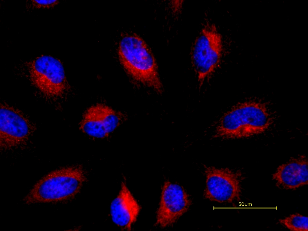
Cell Navigator® TMR Ceramide Golgi Staining Kit
Red Fluorescence
The Golgi apparatus is a complex of vesicles and folded membranes within the cytoplasm of most eukaryotic cells, involved in secretion and intracellular transport. It modifies proteins and lipids that have been built in the endoplasmic reticulum (ER) and prepares them for export outside of the cell. It also plays a significant role in the transport of lipids throughout the cell and the formation of lysosomes. Cell Navigator® TMR Ceramide Golgi Staining kit provides a simple and rapid way to stain Golgi in red fluorescence in live cells. Golgi apparatus is stained through the formation of the respective fluorescent metabolites. Cell Navigator® TMR Ceramide Golgi Staining Kit provides an optimized assay method for examining the morphology of the Golgi apparatus with a fluorescence microscope. In addition, GGR169-ceramide is pH-sensitive, thus uniquely serving as an excellent Golgi pH probe too.
| Catalog | Size | Price | Quantity |
|---|---|---|---|
| 22752 | 100 Tests | Price |

Spectral properties
| Excitation (nm) | 544 |
| Emission (nm) | 570 |
Usage and storage
| Intended use | Research Use Only (RUO) |
Instrument settings
| Fluorescence microscope | |
| Excitation | Cy3/TRITC filter set |
| Emission | Cy3/TRITC filter set |
| Recommended plate | Black wall/clear bottom |
Contact us
| Telephone | |
| Fax | |
| sales@aatbio.com | |
| International | See distributors |
| Bulk request | Inquire |
| Custom size | Inquire |
| Technical Support | Contact us |
| Request quotation | Request |
| Purchase order | Send to sales@aatbio.com |
| Shipping | Standard overnight for United States, inquire for international |
Page updated on July 15, 2026