Products
Services
Resources
Selection Guides
About
Cyanine 3 monosuccinimidyl ester
equivalent to Cy3® NHS ester
Cyanine 3 monosuccinimidyl ester is an amine-reactive orange fluorescent dye that forms stable amide bonds with proteins and antibodies, widely used for creating bright, photostable bioconjugates for microscopy and flow cytometry.
- Orange fluorescence: Provides bright, photostable signal optimal for 532 nm laser excitation in microscopy and flow cytometry
- Amine-reactive conjugation: Forms stable amide bonds with lysine residues at pH 8.5 for efficient protein and antibody labeling
- Multicolor imaging applications: Compatible with FITC and Cy5 for triple-labeling experiments in immunofluorescence and FISH
| Catalog | Size | Price | Quantity |
|---|---|---|---|
| 141 | 1 mg | Price |

Physical properties
| Molecular weight | 829.03 |
| Solvent | DMSO |
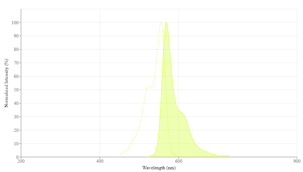
Spectral properties
| Correction factor (260 nm) | 0.07 |
| Correction factor (280 nm) | 0.073 |
| Extinction coefficient (cm -1 M -1) | 150000 1 |
| Excitation (nm) | 555 |
| Emission (nm) | 569 |
| Quantum yield | 0.15 1 |
Usage and storage
| Intended use | Research Use Only (RUO) |
| Storage | Freeze (< -15 °C); Minimize light exposure |
| CAS | 146368-16-3 |
Contact us
| Telephone | |
| Fax | |
| sales@aatbio.com | |
| International | See distributors |
| Bulk request | Inquire |
| Custom size | Inquire |
| Technical Support | Contact us |
| Request quotation | Request |
| Purchase order | Send to sales@aatbio.com |
| Shipping | Standard overnight for United States, inquire for international |
Page updated on September 5, 2024