Products
Services
Resources
Selection Guides
About
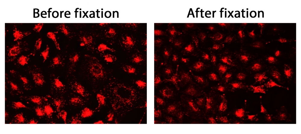
CytoFix™ Red Lysosomal Stain
Lysosomes are cellular organelles which contain acid hydrolase enzymes to break up waste and cellular debris through a process known as autophagy. AAT Bioquest offers CytoFix™ Red lysosomal stain for selectively staining lysosomes. CytoFix™ Red lysosomal stain is well retained in lysosomes even after fixation. The dye permeates intact live cells and gets trapped in lysosomes. The fluorescence in lysosomes generated by this dye is well retained at least for 1 week, making it an excellent lysosomal tracking dye. The key features of this stain are its high staining efficiency, long retention after fixation with minimal hands on time. CytoFix™ Red lysosomal stain can be used with GFP expressed cells or with other organelles stains for multicolor analysis. It can be used for both suspension and adherent cells and readily adapted for a wide variety of fluorescence platforms.
| Catalog | Size | Price | Quantity |
|---|---|---|---|
| 23210 | 500 Tests | Price |

Physical properties
| Solvent | DMSO |
Spectral properties
| Excitation (nm) | 565 |
| Emission (nm) | 585 |
Storage, safety and handling
| H-phrase | H303, H313, H333 |
| Hazard symbol | XN |
| Intended use | Research Use Only (RUO) |
| R-phrase | R20, R21, R22 |
| Storage | Freeze (< -15 °C); Minimize light exposure |
| UNSPSC | 12171501 |
| CAS | N/A |
Instrument settings
| Fluorescence microscope | |
| Excitation | Cy3/TRITC filter set |
| Emission | Cy3/TRITC filter set |
| Recommended plate | Black wall/clear bottom |
| Instrument specification(s) | Cy3/TRITC filter set |
Contact us
| Telephone | |
| Fax | |
| sales@aatbio.com | |
| International | See distributors |
| Bulk request | Inquire |
| Custom size | Inquire |
| Technical Support | Contact us |
| Request quotation | Request |
| Purchase order | Send to sales@aatbio.com |
| Shipping | Standard overnight for United States, inquire for international |
Page updated on July 11, 2026