logo
AAT Bioquest

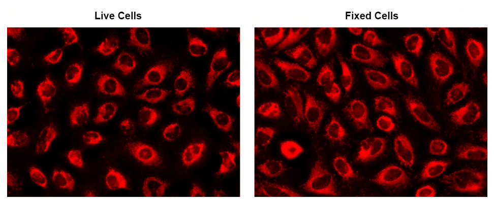
CytoFix™ Red Mitochondrial Stain

The fluorescence images of HeLa cells stained with CytoFix™ MitoRed in a 96-well black-wall clear-bottom plate. Images were acquired before (Left) and after (Right) fixation with 4% formaldehyde solution for 20 minutes at RT. The cells were imaged using a fluorescence microscope equipped with a Cy3/TRITC filter.
Ordering information
Price
Catalog Number
Unit Size
Quantity
Add to cart
Additional ordering information
Telephone1-800-990-8053
Fax1-800-609-2943
Emailsales@aatbio.com
InternationalSee distributors
Bulk requestInquire
Custom sizeInquire
ShippingStandard overnight for United States, inquire for international
Request quotation
Physical properties
Molecular weightN/A
SolventDMSO
Spectral properties
Excitation (nm)564
Emission (nm)651
Storage, safety and handling
H-phraseH303, H313, H333
Hazard symbolXN
Intended useResearch Use Only (RUO)
R-phraseR20, R21, R22
StorageFreeze (< -15 °C); Minimize light exposure
UNSPSC12171501

OverviewpdfSDSpdfProtocol


See also: Mitochondria
CAS
N/A
Molecular weight
N/A
Excitation (nm)
564
Emission (nm)
651
CytoFix™ Red mitochondrial stain is a dye that selectively stains mitochondria independent of mitochondrial membrane potential gradient. Due to this functionality, CytoFix™ Red mitochondrial stain is well retained in mitochondria even after fixation. The dye permeates intact live cells and gets trapped in live cells. Its key features include high staining efficiency, long retention after fixation with minimal hands on time. CytoFix™ Red mitochondrial stain can be used with GFP expressed cells without overlapping the fluorescence of GFP, making it useful for multiplexing analysis. It can be used for both suspension and adherent cells and readily adapted for a wide variety of fluorescence platforms.

Platform


Fluorescence microscope

ExcitationCy3/TRITC filter set
EmissionCy3/TRITC filter set
Recommended plateBlack wall/clear bottom
Instrument specification(s)Cy3/TRITC filter set

Example protocol


AT A GLANCE

  1. Prepare cells in growth medium
  2. Incubate cells with CytoFix™ MitoRed working solution for 20-30 minutes at 37 °C
  3. Remove CytoFix™ MitoRed working solution
  4. Fix cells with formaldehyde (Optional)
  5. Analyze under fluorescence microscope with Cy3/TRITC filter set

CELL PREPARATION

For guidelines on cell sample preparation, please visit https://www.aatbio.com/resources/guides/cell-sample-preparation.html

PREPARATION OF WORKING SOLUTION

Add 20 µL of the stock solution into 10 mL of Hanks and 20 mM Hepes buffer (HHBS) or buffer of your choice or cell culture medium, and mix well.

Note: 20 µL stock solution is enough for one 96-well plate assay. The staining conditions may be modified according to the particular cell type and the permeability of the cells or tissues to the probe.

Note: Unused CytoFix™ MitoRed stock solution can be aliquoted and stored at ≤ -20 °C with smaller aliquots. Protect from light and avoid repeated freeze-thaw cycles.

SAMPLE EXPERIMENTAL PROTOCOL

  1. Prepare cells in growth medium.
  2. Add 100 µL/well (96-well plate) or 50 µL/well (384-well plate) of CytoFix™ MitoRed working solution in the cell plate.

    Note: The optimal concentration of the cell membrane probe varies depending on the specific application.

    Note: Serum in the culture growth medium may interfere with the staining. Staining in the absence of a culture growth medium should improve the staining intensity.

  3. Incubate the cells at 37 °C for 20-30 minutes, protected from light.
  4. Remove working solution in each well. Wash cells with HHBS or buffer of your choice. (Optional)
  5. Optional: Fix the cells with a 4 % solution of paraformaldehyde for 20-30 minutes at room temperature. Wash twice to get rid of the fixation solution.

  6. Observe the fluorescence signal in cells using fluorescence microscope with a Cy3/TRITC filter set.

Spectrum


Open in Advanced Spectrum Viewer
spectrum

Spectral properties

Excitation (nm)564
Emission (nm)651

Product Family


NameExcitation (nm)Emission (nm)
CytoFix™ Red Lysosomal Stain565585

Images