logo
AAT Bioquest

CytoTell™ Green

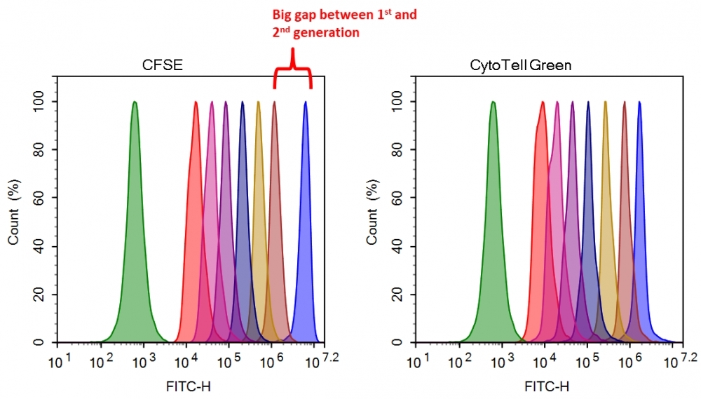
Flow cytometry combined with fluorescence staining is a powerful tool to analyze heterogeneous cell populations. Among all the existing fluorescent dyes CFSE is the preferred cell proliferation indicator that is widely used for live cell analysis. However, there are a few severe problems associated with the use of CFSE for monitoring cell proliferation. 1). CFSE is highly toxic to cells. CFSE indiscriminately reacts with all amino groups, thus changes many critical intracellular protein functions (such as cell membrane GPCRs); 2). CFSE has slow response and is inconvenient to use. The CFSE fluorescence intensity of the 2nd generation cells is decreased more than 10 fold from the 1st generation. You would have to wait for another generation to start the cell proliferation analysis. 3). Medium removal is required. You would have to remove medium for cell analysis with a flow cytometer since CFSE reacts with medium components. CytoTell™ Green is developed to eliminate these limitations. It has distinct advantages over CFSE. 1). CytoTell™ Green has very minimal cytotoxicity compared to CFSE. It is well retained in cells since the probe is designed to minimize the MDR interaction that generally pumps many cell stains out cells; 2). CytoTell™ Green exhibits much faster response and is more convenient to use than CFSE. There is no fluorescence intensity gap between 1st and 2nd generation of cells. As cells divide, CytoTell™ Green is distributed equally between daughter cells that can be measured as successive halving of the fluorescence intensity of the dye; 3). There is no need to remove medium since CytoTell™ Green does not react with medium components; 4). CytoTell™ Green is more sensitive than CFSE. Up to 9 generations may be visualized; 5). CytoTell™ Green is much more stable than CFSE. CytoTell™ Green stock solution can be stored at room temperature for a few days. CytoTell™ Green can also be used for long term tracking of labeled cells. Analysis using two-parameter plots may provide better resolution of each generation, especially between undivided cells and the first generation. Cells labeled with CytoTell™ Green may be fixed and permeabilized for analysis of intracellular targets using standard formaldehyde-containing fixatives and saponin-based permeabilization buffers. CytoTell™ Green has a peak excitation of 510 nm and can be excited by the blue (488 nm) laser line, making it compatible with FITC filter set.
Example protocol

AT A GLANCE

Protocol Summary
  1. Prepare cells with test compounds
  2. Add 1X dye working solution
  3. Incubate dyes with cells at room temperature or 37 oC for 10 to 30 minutes
  4. Remove the dye working solution
  5. Analyse with flow cytometer with appropriate filter set
Important Note

Bring all the kit components to room temperature before starting the experiment.

Note: The CytoTell™ dyes are lyophilized powders. They should be stable for at least 6 months if stored at -20 °C, protecting from light and avoiding freeze/thaw cycles.

PREPARATION OF STOCK SOLUTIONS

Unless otherwise noted, all unused stock solutions should be divided into single-use aliquots and stored at -20 °C after preparation. Avoid repeated freeze-thaw cycles

CytoTell™ dye stock solution (500X)

Add 500 µL DMSO into the dye powder vial, and mix it well by vortexing to have a stock solution (500X).

Note: The stock solution should be used promptly. Any remaining solution should be aliquoted and frozen at < - 20 oC. Avoid repeated freeze-thaw cycles, and protect from light.

PREPARATION OF WORKING SOLUTION

CytoTell™ dye working solution (1X)

Dilute the 500X DMSO stock solution at 1 to 500 in Hanks and 20 mM Hepes buffer (HHBS) or the buffer of your choice, pH 7 (such as 1 µL of 500X DMSO stock solution to 500 µL buffer) right before use. Mix them well by vortexing.

Note: The final concentration of the dye working solution should be empirically determined for different cell types and/or experimental conditions. It is recommended to test at the concentrations that are at least over tenfold range. Such as, CytoTell™ Red might use much less amount in some cell types than the recommended concentrations.

SAMPLE EXPERIMENTAL PROTOCOL

  1. Treat cells with test compounds for a desired period of time.
  2. Centrifuge the cells to get 1-5 × 10⁵ cells per tube.
  3. Resuspend cells in 500 µL of the CytoTell™ dye working solution. Optional: One can add the 500X DMSO stock solution into the cells directly without medium removing (such as, add 1 µL500X DMSO stock solution into 500 µL cells)
  4. Incubate cells with a dye solution at room temperature or 37 °C for 10 to 30 minutes, protected from light.
  5. Remove the dye working solution from the cells, wash the cells with HHBS or buffer of your choice. Resuspend cells in 500 µL of pre-warmed HHBS or medium to get 1-5 × 10⁵ cells per tube.
  6. Monitor the fluorescence change at respected Ex/Em (see Key Parameters) with a flow cytometer or a fluorescence microscope.

Calculators

Common stock solution preparation

Table 1. Volume of DMSO needed to reconstitute specific mass of CytoTell™ Green to given concentration. Note that volume is only for preparing stock solution. Refer to sample experimental protocol for appropriate experimental/physiological buffers.

0.1 mg0.5 mg1 mg5 mg10 mg
1 mM238.527 µL1.193 mL2.385 mL11.926 mL23.853 mL
5 mM47.705 µL238.527 µL477.054 µL2.385 mL4.771 mL
10 mM23.853 µL119.263 µL238.527 µL1.193 mL2.385 mL

Molarity calculator

Enter any two values (mass, volume, concentration) to calculate the third.

Mass (Calculate)Molecular weightVolume (Calculate)Concentration (Calculate)Moles
/=x=

Spectrum

Product family

NameExcitation (nm)Emission (nm)
CytoTell™ Blue410445
CytoTell™ Orange541560

Citations

View all 26 citations: Citation Explorer
The CH1 domain influences the expression and antigen sensing of the HIV-specific CH31 IgM-BCR and IgG-BCR
Authors: Ortiz, Yaneth and Anasti, Kara and Pane, Advaiti K and Cronin, Kenneth and Alam, S Munir and Reth, Michael
Journal: Proceedings of the National Academy of Sciences (2024): e2404728121
A co-culture system of macrophages with breast cancer tumoroids to study cell interactions and therapeutic responses
Authors: Raffo-Romero, Antonella and Ziane-Chaouche, Lydia and Salom{\'e}-Desnoulez, Sophie and Hajjaji, Nawale and Fournier, Isabelle and Salzet, Michel and Duhamel, Marie
Journal: Cell Reports Methods (2024)
Optimizing lentiviral vector formulation conditions for efficient ex vivo transduction of primary human T cells in chimeric antigen receptor T-cell manufacturing
Authors: Luostarinen, Annu and Kailaanm{\"a}ki, Anssi and Turkki, Vesa and K{\"o}ylij{\"a}rvi, Marjut and K{\"a}yhty, Piia and Leinonen, Hanna and Albers-Skirdenko, Vita and Lipponen, Eevi and Yl{\"a}-Herttuala, Seppo and Kaartinen, Tanja and others,
Journal: Cytotherapy (2024)
Effective targeting of breast cancer by the inhibition of P-glycoprotein mediated removal of toxic lipid peroxidation byproducts from drug tolerant persister cells
Authors: Szeb{\'e}nyi, Korn{\'e}lia and F{\"u}redi, Andr{\'a}s and Bajtai, Eszter and Sama, Sai Nagender and Csiszar, Agnes and Gombos, Bal{\'a}zs and Szab{\'o}, P{\'a}l and Grusch, Michael and Szak{\'a}cs, Gergely
Journal: Drug Resistance Updates (2023): 101007
Concurrent stimulation of monocytes with CSF1 and polarizing cytokines reveals phenotypic and functional differences with classical polarized macrophages
Authors: An, Liying and Michaeli, Julia and Pallavi, Prama and Breedijk, Annette and Xu, Xin and Dietrich, Nadine and Sigl, Martin and Keese, Michael and Nitschke, Katja and Jarczyk, Jonas and others,
Journal: Journal of Leukocyte Biology (2022)
Page updated on September 19, 2024

Ordering information

Price
Unit size
500 Tests
2x500 Tests
Catalog Number
Quantity
Add to cart

Additional ordering information

Telephone1-800-990-8053
Fax1-800-609-2943
Emailsales@aatbio.com
InternationalSee distributors
Bulk requestInquire
Custom sizeInquire
Technical SupportContact us
Purchase orderSend to sales@aatbio.com
ShippingStandard overnight for United States, inquire for international
Request quotation

Physical properties

Molecular weight

419.24

Solvent

DMSO

Spectral properties

Excitation (nm)

510

Emission (nm)

525

Storage, safety and handling

H-phraseH303, H313, H333
Hazard symbolXN
Intended useResearch Use Only (RUO)
R-phraseR20, R21, R22

Storage

Freeze (< -15 °C); Minimize light exposure
UNSPSC12352200

Platform

Flow cytometer

Excitation488 nm laser
Emission530, 30 nm filter
Instrument specification(s)FITC channel
Cell proliferation assay with CytoTell&trade; Green and CFSE.&nbsp; Jurkat cells (~2&times;10<sup>6</sup>&nbsp;cells/mL) were stained with CytoTell&trade; Green or CFSE on Day 0. The cells were passed serially at 1:1 ratio on the day specified. Fluorescence intensity was measured with ACEA NovoCyte 3000 flow cytometer in&nbsp;FITC channel on the day of passage. Successive generations were represented by different colors.