logo
Products
Technologies
Applications
Services
Resources
Selection Guides
About
DAPI
4,6-Diamidino-2-phenylindole, dihydrochloride; 10 mM solution in water
DAPI is a fluorescent stain that binds strongly to DNA. It is used extensively in fluorescence microscopy. Since DAPI passes through an intact cell membrane, it can be used to stain live cells besides fixed cells. For fluorescence microscopy, DAPI is excited with ultraviolet light. When bound to double-stranded DNA its absorption maximum is at 358 nm and its emission maximum is at 461 nm. One drawback of DAPI is that its emission is fairly broad. DAPI also binds to RNA although it is not as strongly fluorescent as it binds to DNA. Its emission shifts to around 500 nm when bound to RNA. DAPI's blue emission is convenient for multiplexing assays since there is very little fluorescence overlap between DAPI and green-fluorescent molecules like fluorescein and green fluorescent protein (GFP), or red-fluorescent stains like Texas Red. Besides labeling cell nuclei, DAPI is also used for the detection of mycoplasma or virus DNA in cell cultures.
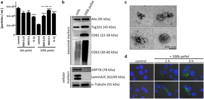
<em>L. pneumophila</em> infection increases the secretion of EVs in THP-1 cells. (a) Amount of EVs in response to <em>L. pneumophila</em> infection. THP-1 cells were treated with IL-1&beta; (1&thinsp;ng/mL) or infected with <em>L. pneumophila</em> (MOI 0.25 or 0.5, respectively) for 24&thinsp;h. NTA was performed with the distinct particle fractions separated by differential centrifugation. (b) Western blot for exosomal marker proteins. Whole cell lysate or 100&thinsp;k pellet derived from uninfected THP-1 cells were used. Equal protein amounts were loaded. (c) Transmission electron microscopy with 100&thinsp;k pellet. Purified EVs from THP-1 cells were fixed and visualized after negative staining with uranyl acetate. Scale bar represents 100&thinsp;nm. (d) Uptake of A549-derived EVs by THP-1 cells. A549 cells were stained with the membrane dye PKH67. EVs were collected (100&thinsp;k pellet) and incubated with THP-1 cells for 1 or 3&thinsp;h, respectively. Nuclei were stained by DAPI and pictures were taken with an original magnification of 630x. A: Data are shown as mean&thinsp;+&thinsp;SEM of three independent experiments. **p&thinsp;&lt;&thinsp;0.01, ***p&thinsp;&lt;&thinsp;0.001, ****p&thinsp;&lt;&thinsp;0.0001. B&ndash;D: representative results. Source: <strong><em>Legionella pneumophila</em> infection activates bystander cells differentially by bacterial and host cell vesicles </strong>by Jung et al., <em>Scientific Reports,</em> July 2017.
CatalogSize
Price
Quantity
175072 mL
Price
 
Physical properties

Molecular weight350.25
SolventWater
Spectral properties

Extinction coefficient (cm -1 M -1)
27000
Excitation (nm)359
Emission (nm)457
Storage, safety and handling

Certificate of OriginDownload PDF
H-phraseH303, H313, H340
Hazard symbolT
Intended useResearch Use Only (RUO)
R-phraseR20, R21, R68
StorageFreeze (< -15 °C); Minimize light exposure
UNSPSC41116134
CAS28718-90-3
Contact us

Telephone
Fax
Emailsales@aatbio.com
InternationalSee distributors
Bulk requestInquire
Custom sizeInquire
Technical SupportContact us
Request quotationRequest
Purchase orderSend to sales@aatbio.com
ShippingStandard overnight for United States, inquire for international
Page updated on September 19, 2024