logo
AAT Bioquest

FerroBrite™ Green

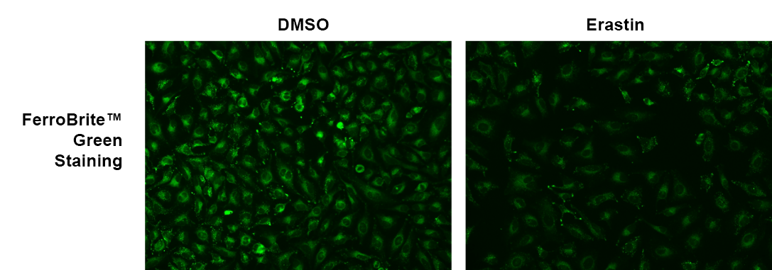
Ferroptosis is an iron-dependent form of regulated cell death associated with the increase in lipid peroxides. Divalent iron (Fe2+) can lead to spontaneous lipid peroxidation through the Fenton reaction. Ferroptosis is regulated by signaling pathways that control iron storage and oxidative stress. FerroBrite™ Green has been developed for detecting ferroptosis via fluorescence imaging. FerroBrite™ Green has superior photostability and responds quickly to ferroptosis. The dye is permeable to live cells. Upon induction of ferroptosis, the imbalance of Fe2+ causes the reduction in the fluorescence intensity of FerroBrite™ Green. The fluorescence of FerroBrite™ Green can be readily monitored using the common FITC filter, which is equipped in most of fluorescence instruments. FerroBrite™ Green enables the real-time tracking of ferroptosis.
Example protocol

AT A GLANCE

Important Note

Before initial use, thaw FerroBrite™ Green at room temperature and briefly centrifuge to collect the dried pellet.

Subsection title
  1. Treat the cells as desired.

  2. Remove the treatment and add 100 µL of FerroBrite™ Green working solution.

  3. Incubate at 37 °C for 30 minutes.

  4. Use an Ex/Em = 450/550 filter set to monitor the fluorescence signal.

PREPARATION OF STOCK SOLUTIONS

Unless otherwise noted, all unused stock solutions should be divided into single-use aliquots and stored at -20 °C after preparation. Avoid repeated freeze-thaw cycles

FerroBrite™ Green Stock Solution
  1. Prepare a 5 to 10 mM FerroBrite™ Green stock solution in DMSO. For example, to make a 10 mM FerroBrite™ Green stock solution add 284 µL of DMSO to the vial of FerroBrite™ Green, and mix thoroughly.

    Note: Prepare single-use aliquots of any remaining FerroBrite™ Green stock solution. Store aliquots at ≤ -20 º C, protected from light. Avoid freeze/thaw cycles.

PREPARATION OF WORKING SOLUTION

FerroBrite™ Green Working Solution
  1. Prepare a 10 to 20 μM working solution by diluting the FerroBrite™ Green stock solution into Hanks' solution with 20 mM Hepes buffer (HHBS, AAT Cat No. 20011).

    Note: For optimal results, use this solution within 2 hours of preparation.

    Note: Protect the working solution from light by covering it with foil or placing it in the dark.

SAMPLE EXPERIMENTAL PROTOCOL

  1. Plate the cells as desired in a 96-well black wall-clear bottom plate.

  2. Add the drug of interest to the cells at the necessary concentration.

    Note: For a positive control, add Erastin at a concentration of 10 µM to the cells. Incubate the cells with Erastin for 5 to 6 hours at 37°C in a 5% CO2 incubator to induce ferroptosis.

  3. Remove the cell culture medium and add 100 µL of FerroBrite™ Green working solution to the cells.

  4. Incubate the cells at 37°C for 20 to 30 minutes, keeping them protected from light.

    Note: The optimal concentration and incubation time for FerroBrite™ Green may vary depending on the cell line used. It is recommended to test various concentrations to determine the most effective dose.

  5. Remove the FerroBrite™ Green working solution and wash the cells twice using HHBS buffer.

  6. Add HHBS buffer and analyze the cells using a fluorescence microscope with an Ex/Em = 450/550 nm filter set.

Spectrum

References

View all 50 references: Citation Explorer
A dual-labeling fluorescent probe to track lysosomal polarity and endoplasmic reticulum dynamics during ferroptosis.
Authors: Zhao, Zhao and Jin, Wendong and Wu, Mengfan and Lin, Qingyu and Duan, Yixiang
Journal: Chemical communications (Cambridge, England) (2024): 7773-7776
Rational design of a lysosome-targeted fluorescent probe for monitoring the generation of hydroxyl radicals in ferroptosis pathways.
Authors: Zhong, Lili and Fu, Datian and Xu, Jin and Tan, Linyan and Wu, Haimei and Wang, Min
Journal: RSC advances (2024): 12864-12872
A sensitive "turn-on" Schiff-base fluorescent probe for the selective detection of Fe3+ and bio-imaging.
Authors: Che, Yiran and Yang, Jingying and Dong, Zhenming and Wang, Jianhua and Yan, Xiaoqing and Wang, Yu and Shuang, Shaomin
Journal: Spectrochimica acta. Part A, Molecular and biomolecular spectroscopy (2024): 123799
A D-π-A-type ratiometric fluorescent probe to detect polarity changes and inhibition effect during ferroptosis.
Authors: Mehmood, Abdul Hadi and Chang, Jia and Wang, Yan and Li, Shijing and Ma, Jiale and Dong, Baoli and Liu, Hong
Journal: Analytical methods : advancing methods and applications (2024): 3486-3491
Peroxynitrite imaging in ferroptosis-mediated drug-induced liver injury with a near-infrared fluorescence probe.
Authors: Liu, Ruixin and Jiang, Haijing and Yang, Wenjie and Zheng, Zhijuan and Wang, Xiaoming and Tian, Zhenhua and Wang, Danyang and Kan, Dongfang and Zhang, Dan and Tang, Zhixin
Journal: Analytica chimica acta (2024): 342673
Page updated on October 11, 2024

Ordering information

Price
Unit size
Catalog Number20205
Quantity
Add to cart

Additional ordering information

Telephone1-800-990-8053
Fax1-800-609-2943
Emailsales@aatbio.com
InternationalSee distributors
Bulk requestInquire
Custom sizeInquire
Technical SupportContact us
Purchase orderSend to sales@aatbio.com
ShippingStandard overnight for United States, inquire for international
Request quotation

Physical properties

Molecular weight

352.45

Solvent

DMSO

Spectral properties

Excitation (nm)

453

Emission (nm)

552

Storage, safety and handling

Certificate of OriginDownload PDF
H-phraseH303, H313, H333
Hazard symbolXN
Intended useResearch Use Only (RUO)
R-phraseR20, R21, R22

Storage

Freeze (< -15 °C); Minimize light exposure

Platform

Fluorescence microscope

Excitation450 nm
Emission550 nm
Recommended plateBlack wall, clear bottom
Erastin induced ferroptosis in Hela cells. The fluorescence response of FerroBrite™ Green (20 µM) was evaluated in HeLa cells, in the absence and presence of 6-hour Erastin treatment at 37°C, 5% CO2 incubator. Fluorescence intensities were measured using fluorescence microscopy equipped with a FITC filter.