logo
AAT Bioquest

FluoroQuest™ Fluorescence Quantum Yield Determination Kit *Optimized for Bioconjugates*

Principle of fluorescence. Electrons are excited to a higher energy level by external source. Upon return to their ground state, a set quanta of photons are release proportional to the energy loss by electrons. This release of photons represents the fluorescence emission.The fluorophore's quantum yield is the ratio of its emitted photons to the photons it absorbed.
Ordering information
Price
Catalog Number
Unit Size
Quantity
Add to cart
Additional ordering information
Telephone1-800-990-8053
Fax1-800-609-2943
Emailsales@aatbio.com
InternationalSee distributors
Bulk requestInquire
Custom sizeInquire
ShippingStandard overnight for United States, inquire for international
Request quotation
Storage, safety and handling
H-phraseH303, H313, H333
Hazard symbolXN
Intended useResearch Use Only (RUO)
R-phraseR20, R21, R22
UNSPSC12171501

OverviewpdfSDSpdfProtocol


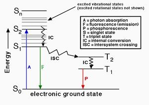
When a fluorophore absorbs a photon of light, an energetically excited state is formed. The fate of this species is varied, depending upon the exact nature of the fluorophore and its surroundings, but the end result is deactivation (loss of energy) and return to the ground state. The main deactivation processes which occur are fluorescence (loss of energy by emission of a photon), internal conversion and vibrational relaxation (non-radiative loss of energy as heat to the surroundings), and intersystem crossing to the triplet manifold and subsequent non-radiative deactivation. The fluorescence quantum yield is the ratio of photons absorbed to photons emitted through fluorescence. In other words the quantum yield gives the probability of the excited state being deactivated by fluorescence rather than by another, non-radiative mechanism. The kit provides all the essential components for fluorescence quantum yield determination for biological conjugates. It is optimized for determining the fluorescence quantum yields of fluorescent protein conjugates, peptides, nucleotides and nucleic acids.

Platform


Spectrofluorometer

ExcitationSee Table 1
EmissionSee Table 1

Fluorescence microplate reader

ExcitationSee Table 1
EmissionSee Table 1
Recommended plateSolid black

Components


Example protocol


AT A GLANCE

Experimental considerations

The most reliable method for recording fluorescence quantum yield is the comparative method of Williams et al. It involves the use of well characterized standard samples with known fluorescence quantum yield values. Essentially, solutions of the standard and test samples with identical absorbance at the same excitation wavelength can be assumed to be absorbing the same number of photons. Hence, a simple ratio of the integrated fluorescence intensities of the two solutions (recorded under identical conditions) will yield the ratio of the quantum yield values. Since the fluorescence quantum yield for the standard sample (Φs) is known, it is trivial to calculate the fluorescence quantum yield for the sample (Φx).

The standard reference should be chosen to have certain absorbance at the excitation wavelength of choice for the test sample, and, if possible, emit in a similar region to the test sample. Standard 10 mm path length fluorescence cuvettes are sufficient for running the fluorescence measurements. For small amount samples, either a microplate reader or nanodrop device can be used. The absorbance in the 10 mm cuvette should be <0.2. Above this level, non-linear effects may be observed due to inner filter effects, and the resulting quantum yield values may be inaccurate.

This kit is only designed for a one-point quick estimation of the fluorescence quantum yield of a biological conjugate. For more accurate deamination of fluorescence quantum yield there are many factors that have a significant effect on fluorescence quantum yields.

PREPARATION OF STOCK SOLUTION

Unless otherwise noted, all unused stock solutions should be divided into single-use aliquots and stored at -20 °C after preparation. Avoid repeated freeze-thaw cycles.

Make 1 mM stock solutions of the selected standard and test samples using a proper solvent (such as water or DMSO).

Table 1. Kit components with respective quantum yield in water and similar wavelength dyes for reference standard selection.

ComponentsFluorescence
Quantum Yield
in Water (ΦValue)
Conjugates Labeled with the Following Dyes or Dyes of Similar Wavelength (for Reference Standard Selection)
Reference A 0.98FAM, 6-TET, 6-HEX, 6-JOE, FITC, Cy®2, Alexa Fluor® 488 and 514, iFluor™ 488 and 514, DyLight™ 488, or other dyes that have an emission of 500 ± 50 nm
Reference B 0.20Cy3®, Alexa Fluor® 514, 532, 546 and 555, iFluor™ 514, 532 and 555, DyLight™ 555, TRITC, or other dyes that have an emission of 550 ± 50 nm
Reference C 0.44Texas Red®, Texas Red®-X, Alexa Fluor® 594, iFluor™ 594, California Red™, DyLight™ 594, or other dyes that have an emission of 600 ± 50 nm
Reference D 0.24Cy5®, Cy5.5®, Cy7®, Alexa Fluor® 633, 647, 700 and 750, iFluor™ 633, 647, 700 and 750, DyLight™ 650, 680 and 755, or other dyes that have an emission of 650 ± 50 nm 

SAMPLE EXPERIMENTAL PROTOCOL

  1. Record the absorbance spectra of the selected standard and test samples (0.1 to 5 µM) using the diluted solutions prepared from their stock solutions with an aqueous buffer (such as PBS or TRIS).

  2. Select the excitation wavelength based on the absorption maxima. We recommend to use this following formula to select the desired excitation wavelength:
         Desired excitation(nm) = shorter absorption maximum* – 50nm
    *Selected from either the reference standard or test sample. For example, if your test sample has absorption maximum at 500 nm, and the selected reference standard has the absorption maximum at 490 nm, you should select 440 nm (490 nm  - 50 nm) as excitation wavelength. 

  3. Adjust the concentrations of the selected reference standard and test samples to have the same absorbance (between 0.2 to 0.8) at the selected excitation wavelength. Note: It might be easier to choose the isosbestic point of the reference standard and test sample absorption spectra.

  4. Dilute both the selected reference standard and test sample by 10 times (must be diluted by the same proportion) using the same buffer or pure water.

  5. Record the fluorescence spectra of the same diluted solutions in the 10 mm fluorescence cuvette using a fluorescence spectrophotometer or a microplate reader that can generate corrected fluorescence spectrum. Note: All the fluorescence spectra must be recorded with the same instrument settings. Changing instrument settings between samples will invalidate the quantum yield measurement.

Images


References


View all 14 references: Citation Explorer
Stability and fluorescence quantum yield of CdSe-ZnS quantum dots--influence of the thickness of the ZnS shell
Authors: Grabolle M, Ziegler J, Merkulov A, Nann T, Resch-Genger U.
Journal: Ann N Y Acad Sci (2008): 235
Design of high-affinity peptide conjugates with optimized fluorescence quantum yield as markers for small peptide transporter PEPT1 (SLC15A1)
Authors: Bahadduri PM, Ray A, Kh and elwal A, Swaan PW.
Journal: Bioorg Med Chem Lett (2008): 2555
Fluorescence spectra and fluorescence quantum yield of sulfosalicylic acid
Authors: Wei YJ, Li N, Qin SJ.
Journal: Guang Pu Xue Yu Guang Pu Fen Xi (2004): 647
Measurement of fluorophore concentrations and fluorescence quantum yield in tissue-simulating phantoms using three diffusion models of steady-state spatially resolved fluorescence
Authors: Diamond KR, Farrell TJ, Patterson MS.
Journal: Phys Med Biol (2003): 4135
Fluorescence quantum yield determination of pyrimidine (6-4) pyrimidone photoadducts
Authors: Blais J, Douki T, Vigny P, Cadet J.
Journal: Photochem Photobiol (1994): 402
A comparison of three photosensitizers with respect to efficiency of cell inactivation, fluorescence quantum yield and DNA strand breaks
Authors: Kvam E, Moan J.
Journal: Photochem Photobiol (1990): 769
The fluorescence quantum yield of vitamin A2
Authors: Tsin AT, Pedrozo-Fern and ez HA, Gallas JM, Chambers JP.
Journal: Life Sci (1988): 1379
Fluorescence decay and quantum yield characteristics of acridine orange and proflavine bound to DNA
Authors: Kubota Y, Steiner RF.
Journal: Biophys Chem (1977): 279
Dependence of the fluorescence quantum yield of complexes of acriflavine with tRNA on its structure
Authors: Borisova OF, Potapov AP, Surovaya AN, Trubitsin SN, Vol'kenshtein MV.
Journal: Mol Biol (1974): 411
5-dimethylaminonaphthalene-1-sulfonic acid (DANS acid) as standard for quantum yield of fluorescence
Authors: Himel CM, Mayer RT.
Journal: Anal Chem (1970): 130