logo
AAT Bioquest

FluoroQuest™ PLUS Antifade Mounting Medium

Antifade properties were measured using cells stained with fluorescently labeled secondary antibodies, as listed in the graph. The antifade capability of the VectaShield and FluoroQuest™ PLUS Antifade mounting mediums was measured using a 20X objective with real-time imaging over 10 sec of continuous exposure to the excitation illumination. The intensity after 10 seconds of exposure is expressed as 100% percent of the intensity at zero time. The data shown above in the graph represents the % fluorescence remaining after 3 minutes of continuous exposure.
Ordering information
Price
Catalog Number
Unit Size
Quantity
Add to cart
Additional ordering information
Telephone1-800-990-8053
Fax1-800-609-2943
Emailsales@aatbio.com
InternationalSee distributors
Bulk requestInquire
Custom sizeInquire
ShippingStandard overnight for United States, inquire for international
Request quotation
Storage, safety and handling
H-phraseH303, H313, H333
Hazard symbolXN
Intended useResearch Use Only (RUO)
R-phraseR20, R21, R22
StorageRefrigerated (2-8 °C); Minimize light exposure

OverviewpdfSDSpdfProtocol


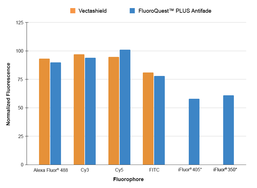
FluoroQuest™ PLUS Antifade Mounting Medium, the latest breakthrough in non-setting media technology, provides unparalleled protection against fading throughout the visible spectrum, including far-red wavelengths. Its compatibility with most commercially available fluorophores makes it a versatile solution for various fluorescence imaging applications. With its improved formulation, it outperforms its predecessor, the FluoroQuest™ Antifade Mounting Medium, by exhibiting no inherent background or toning while ensuring superior retention of fluorophore signals across the spectrum, including far-red wavelengths. Additionally, this superior mounting medium enables mounted sections to be viewed in just one hour, while its non-setting formulation ensures consistent and artifact-free results.

Example protocol


AT A GLANCE

Protocol Summary
  1. Prepare Samples (slides or microplate wells).

  2. Add a drop of a component and mount.

  3. Examine the specimen under microscope.

Important Note

These ready-to-use anti-fading reagents can be applied directly to the washed specimen. Although the reagents have been tested with lots of fixed samples, their optimal anti-fading efficiencies strongly depend on the properties of your samples. We suggest that you try more than one component for your imaging samples to get the ideal component. For example, one component may be more compatible with a fluorescently labeled antibody conjugate (or an enzyme substrate or special mounting specimens that contain lipophilic plasma membrane stains like DiI) than another one.

CELL PREPARATION

For guidelines on cell sample preparation, please visit:

https://www.aatbio.com/resources/guides/cell-sample-preparation.html

SAMPLE EXPERIMENTAL PROTOCOL

  1. Thaw the vial of FluoroQuest™ PLUS Antifade Mounting Medium at room temperature, protected from light.

  2. Remove any excess liquid from your specimen. Add a small drop of the selected component to the specimen. If the sample is on a slide or tissue culture dish, carefully place a coverslip on the drop, avoiding air bubbles. If the sample is on a coverslip, invert the coverslip on a clean glass slide. Remove any excess anti-fading component.

  3. The anti-fading reagents should be incubated for 1 hour to overnight. For long-term storage, seal the coverslip to the slide with nail polish or a plastic sealant. Mounted slides should be stored at 4°C in the dark for optimum sample longevity. The fluorescence imaging would remain stable for many weeks. Samples can be imaged immediately after mounting.

Images