logo
AAT Bioquest

Gelite™ Green Nucleic Acid Gel Staining Kit

OverviewpdfSDSpdfProtocol


Gelite™ Green is a sensitive fluorescent nucleic acid gel stain for detecting nucleic acids in agarose and polyacrylamide gels. Gelite™ Green stain exhibits exceptional affinity for DNA and a large fluorescence enhancement upon binding to DNA, at least an order of magnitude greater than that of ethidium bromide when detected by photography. With a standard 300 nm UV transilluminator and photographic detection, as little as 60 pg dsDNA per band can be detected with Gelite™ Green stain. Gelite™ Green nucleic acid gel stain is nearly two orders of magnitude more sensitive than ethidium bromide for staining oligonucleotides in gels. Our Gelite™ Green Nucleic Acid Gel Staining Gel Kit includes our Gelite™ Green nucleic acid stain with an optimized and robust protocol. It provides a convenient solution for staining nucleic acid samples in gels.
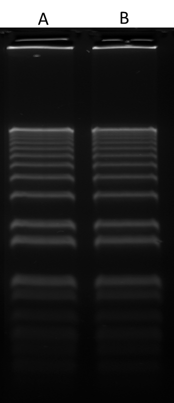
Platform


Transilluminator

Excitation254 nm or 300 nm
EmissionLong path green filter (ex. SYBR or GelStar)

Components


Example protocol


PREPARATION OF WORKING SOLUTION

Add 1 μL of Gelite™ Green Stain (Component A) into 200 μL of 5X Gel Loading Buffer (Component B) to make Gelite™ Green working solution. Protect Gelite™ Green working solution from light by covering it with foil or placing it in the dark.

SAMPLE EXPERIMENTAL PROTOCOL

  1. Prepare DNA samples as you desired.
  2. Add 4 µL of Gelite™ Green working solution into 16 µL of DNA samples and mix well. Incubate at room temperature for 5 - 15 minutes prior to electrophoresis.
  3. Run gels based on your standard protocol.
  4. Image the gel with a 300 nm ultraviolet or 254 nm transilluminator, or a laser-based gel scanner using a long path green filter such as a SYBR® filter or GelStar® filter. 

Images


References


View all 22 references: Citation Explorer
Plasmalemma permeability and necrotic cell death phenotypes after intracerebral hemorrhage in mice
Authors: Zhu X, Tao L, Tejima-M and eville E, Qiu J, Park J, Garber K, Ericsson M, Lo EH, Whalen MJ.
Journal: Stroke (2012): 524
Intra-organ Biodistribution of Gold Nanoparticles Using Intrinsic Two-photon Induced Photoluminescence
Authors: Park J, Estrada A, Schwartz JA, Diagaradjane P, Krishnan S, Dunn AK, Tunnell JW.
Journal: Lasers Surg Med (2010): 630
Screening by imaging: scaling up single-DNA-molecule analysis with a novel parabolic VA-TIRF reflector and noise-reduction techniques
Authors: van 't Hoff M, Reuter M, Dryden DT, Oheim M.
Journal: Phys Chem Chem Phys (2009): 7713
Novel anatomic structures in the brain and spinal cord of rabbit that may belong to the Bonghan system of potential acupuncture meridians
Authors: Lee BC, Kim S, Soh KS.
Journal: J Acupunct Meridian Stud (2008): 29
Triplet fraction buildup effect of the DNA-YOYO complex studied with fluorescence correlation spectroscopy
Authors: Shimizu M, Sasaki S, Kinjo M.
Journal: Anal Biochem (2007): 87
DNA length evaluation using cyanine dye and fluorescence correlation spectroscopy
Authors: Shimizu M, Sasaki S, Tsuruoka M.
Journal: Biomacromolecules (2005): 2703
Implementation of accurate and fast DNA cytometry by confocal microscopy in 3D
Authors: Ploeger LS, Huisman A, van der Gugten J, van der Giezen DM, Belien JA, Abbaker AY, Dullens HF, Grizzle W, Poulin NM, Meijer GA, van Diest PJ.
Journal: Cell Oncol (2005): 225
TO-PRO-3 is an optimal fluorescent dye for nuclear counterstaining in dual-colour FISH on paraffin sections
Authors: Bink K, Walch A, Feuchtinger A, Eisenmann H, Hutzler P, Hofler H, Werner M.
Journal: Histochem Cell Biol (2001): 293
Oxazole yellow homodimer YOYO-1-labeled DNA: a fluorescent complex that can be used to assess structural changes in DNA following formation and cellular delivery of cationic lipid DNA complexes
Authors: Wong M, Kong S, Dragowska WH, Bally MB.
Journal: Biochim Biophys Acta (2001): 61
Photophysical properties of fluorescent DNA-dyes bound to single- and double-stranded DNA in aqueous buffered solution
Authors: Cosa G, Focsaneanu KS, McLean JR, McNamee JP, Scaiano JC.
Journal: Photochem Photobiol (2001): 585