Products
Services
Resources
Selection Guides
About
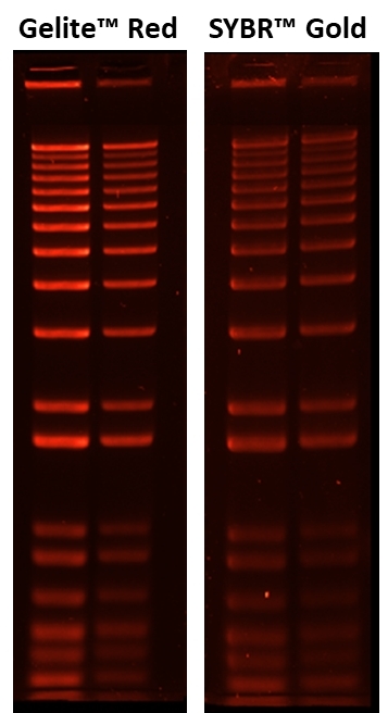
Gelite™ Red Nucleic Acid Gel Stain
Replaced by #17706
Gelite™ Red is the most recent addition to our Gelite™ nucleic acid gel stain family. Now we have a complete family of multicolor gel stains for detecting nucleic acids in gels. Gelite™ Red is an extremely sensitive nucleic acid gel stain for detecting DNA in gels using a standard 300 nm UV transilluminator and Polaroid 667 black-and-white print film. Under the same conditions it is more sensitive than the popular SYBR® Gold gel stain. This remarkable sensitivity can be attributed to a combination of unique dye characteristics of Gelite™ Red. Because the nucleic acid“bound Gelite™ Red dye exhibits excitation maxima close to 488 nm and ~300 nm (the emission maximum is ~610 nm), it is compatible with a wide variety of instrumentation, ranging from UV epi- and transilluminators and blue-light transilluminators, to mercury-arc lamp“ and argon-ion laser“based gel scanners. Our Gelite™ Red is a superior alternative to SYBR® Gold, SYBR® Safe and Ethidium Bromide. It provides a convenient solution for staining nucleic acid samples in gels.
| Catalog | Size | Price | Quantity |
|---|---|---|---|
| 17600 | 1 mL | Price |
Storage, safety and handling
| H-phrase | H303, H313, H340 |
| Hazard symbol | T |
| Intended use | Research Use Only (RUO) |
| R-phrase | R20, R21, R68 |
| Storage | Freeze (< -15 °C); Minimize light exposure |
| UNSPSC | 41116134 |
Contact us
| Telephone | |
| Fax | |
| sales@aatbio.com | |
| International | See distributors |
| Bulk request | Inquire |
| Custom size | Inquire |
| Technical Support | Contact us |
| Request quotation | Request |
| Purchase order | Send to sales@aatbio.com |
| Shipping | Standard overnight for United States, inquire for international |
Page updated on July 12, 2023