Products
Technologies
Applications
Services
Resources
Selection Guides
About
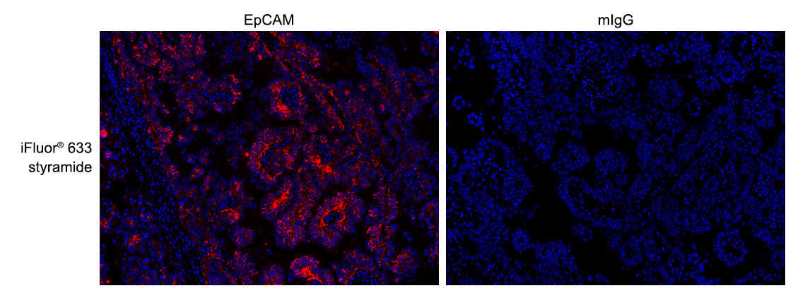
iFluor® 633 Styramide
Superior Replacement for Opal 650
Power Styramide™ Signal Amplification (PSA™) system is one of the most sensitive methods that can detect extremely low-abundance targets in cells and tissues with improved fluorescence signal 10-50 times higher than the widely used tyramide (TSA) reagents. In combination with our superior iFluor® dyes that have higher florescence intensity, increased photostability and enhanced water solubility, the iFluor® dye-labeled Styramide™ conjugates can generate fluorescence signal with significantly higher precision and sensitivity (more than 100 times) than standard ICC/IF/IHC. PSA utilizes the catalytic activity of horseradish peroxidase (HRP) for covalent deposition of fluorophores in situ. PSA radicals have much higher reactivity than tyramide radicals, making the PSA system much faster, more robust and sensitive than the traditional TSA reagents. Compared to tyramide reagents, the Styramide™ conjugates have ability to label the target at higher efficiency and thus generate significantly higher fluorescence signal. Styramide™ conjugates also allow significantly less consumption of primary antibody compared to standard directly conjugate method or tyramide amplification with the same level of sensitivity. iFluor® 633 Styramide is optimized to a superior replacement for Opal Polaris 650 or other spectrally similar fluorescent tyramide conjugates or TSA reagents.
| Catalog | Size | Price | Quantity |
|---|---|---|---|
| 45040 | 100 Slides | Price |

Physical properties
| Molecular weight | 1430.86 |
| Solvent | DMSO |
Spectral properties
| Correction factor (260 nm) | 0.062 |
| Correction factor (280 nm) | 0.044 |
| Extinction coefficient (cm -1 M -1) | 250000 1 |
| Excitation (nm) | 640 |
| Emission (nm) | 654 |
| Quantum yield | 0.29 1 |
Storage, safety and handling
| H-phrase | H303, H313, H333 |
| Hazard symbol | XN |
| Intended use | Research Use Only (RUO) |
| R-phrase | R20, R21, R22 |
| Storage | Freeze (< -15 °C); Minimize light exposure |
| UNSPSC | 12171501 |
Instrument settings
| Fluorescence microscope | |
| Excitation | Cy5 filter set |
| Emission | Cy5 filter set |
| Recommended plate | Black wall/clear bottom |
Documents
Contact us
| Telephone | |
| Fax | |
| sales@aatbio.com | |
| International | See distributors |
| Bulk request | Inquire |
| Custom size | Inquire |
| Technical Support | Contact us |
| Request quotation | Request |
| Purchase order | Send to sales@aatbio.com |
| Shipping | Standard overnight for United States, inquire for international |
Page updated on October 31, 2025