logo
AAT Bioquest

iFluor® Ultra 594 maleimide

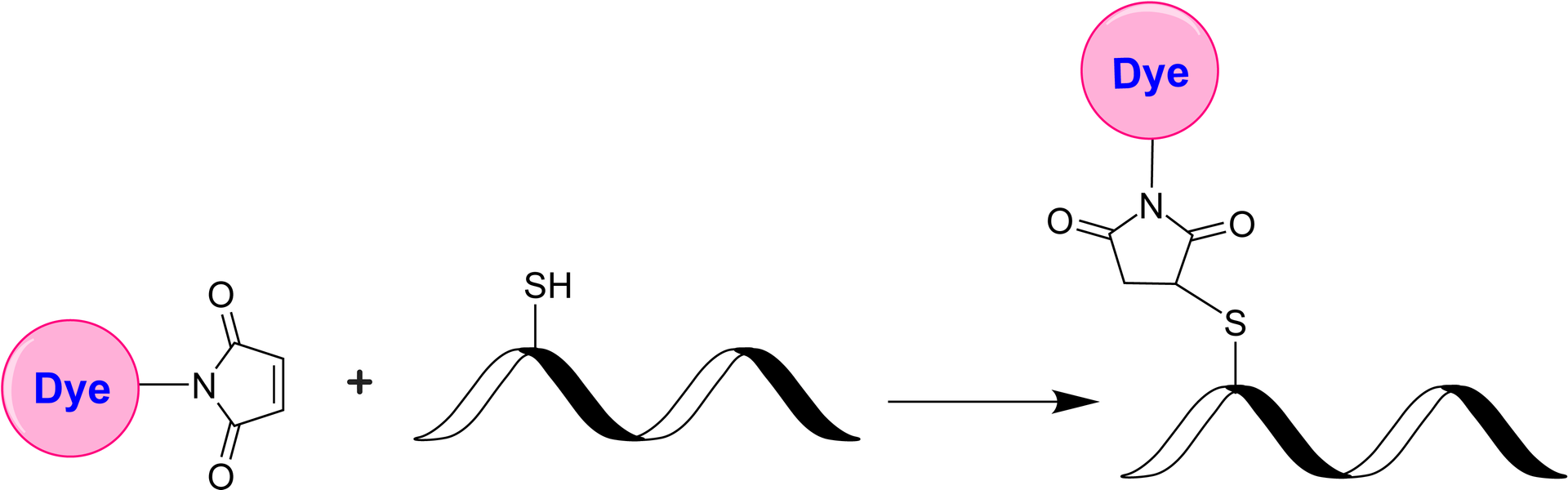
The iFluor® Ultra series represents an enhancement of our established iFluor® dyes, optimized for antibody labeling in fluorescence imaging and flow cytometry. Within this series, iFluor® Ultra 594 is an exceptionally bright red fluorescent dye excitable by 561 nm or 594 nm laser lines, with a peak emission at 601 nm. Antibody conjugates with iFluor® Ultra 594 exhibit superior brightness compared to those labeled with Alexa Fluor® 594 under identical conditions. Additionally, iFluor® Ultra 594 maintains stable fluorescence across a pH range of 4 to 10. The maleimide derivative of iFluor® Ultra 594 is widely used for conjugation to thiol groups on proteins, oligonucleotide thiophosphates, or low molecular weight ligands. Conjugates with iFluor® Ultra 594 demonstrate higher fluorescence intensity and greater photostability compared to those with other spectrally similar fluorophores, enhancing their utility for advanced fluorescence applications. Fluorescent dye-conjugated antibodies are crucial for protein identification in various applications, including fluorescent cell imaging, flow cytometry, western blotting, and immunohistochemistry. The advantages of these conjugates include increased sensitivity, multiplexing capabilities, and ease of use, facilitating complex biological studies.

Example protocol

PREPARATION OF STOCK SOLUTIONS

Unless otherwise noted, all unused stock solutions should be divided into single-use aliquots and stored at -20 °C after preparation. Avoid repeated freeze-thaw cycles

Prepare iFluor® Ultra 594 maleimide stock solution
  1. Allow the vial of iFluor Dye maleimide to warm up to room temperature.
  2. Add anhydrous DMSO to the vial to prepare a 10 mM dye stock solution.
  3. Vortex the vial briefly to fully dissolve the dye, and then centrifuge to collect the dye at the bottom of the vial.
  4. Protect all stock solutions from light as much as possible by wrapping containers in aluminum foil.
Prepare antibody or protein solution for labeling
  1. If your protein already contains a thiol group, prepare the protein at 50-100 uM (for example: 5mg/ml BSA is ~75uM) in 50~100 mM MES buffer or buffers of your choice with pH 6.5~7.0.
  2. If labeling with an intact antibody, reduction of disulfide bonds need to be carried out before maleimide reaction. Prepare antibody in 2-10 mg/ml in a suitable buffer with pH 7.0–7.5. A 10-fold molar excess of a reducing agent such as DTT or TCEP is added to the antibody. If DTT is used, it must be removed by dialysis or desalting to a suitable buffer with pH 6.5~7.0 prior to conjugation. If TCEP is used, it is not necessary to remove excess TCEP during conjugation with maleimides, however, removal of TCEP by dialysis or desalting prior to conjugation gives the better labeling efficiency.

    Below is a sample protocol for generating free thiol groups on antibody:
    1. Prepare 2-10mg/ml IgG solution in PBS.
    2. Prepare a fresh solution of 1 M DTT (15.4 mg/100 µL) in distilled water. 
    3. Add 1- 20 µL of DTT stock per ml of IgG solution while mixing. 
    4. Let the solution stand at room temperature for 30 minutes without additional mixing (to minimize the re-oxidation of cysteines to cystines). 
    5. Pass the reduced IgG over a filtration column pre-equilibrated with 50 mM MES buffer (pH=6.5) to remove excess DTT.
    6. Determine the antibody concentrations. This can be done either spectrophotometrically or colorimetrically.
    7. Carry out the conjugation as soon as possible after this step.

    Note: For the best results, IgG solutions should be > 2 mg/mL.

    Note: The reduction can be carried out in almost any buffer from pH 7 to 7.5, e.g., MES, phosphate, or TRIS buffers.

    Note: Steps 5 can be replaced by dialysis.

     
  3. If your protein doesn’t have a free thiol group or disulfide bond to reduce, a thiolation modification need to be carried out before maleimide conjugation (for example:  using 2-Iminothiolane or 2-IT) to introduce sulfhydryl (-SH) groups to the original amino groups on protein.

SAMPLE EXPERIMENTAL PROTOCOL

This labeling protocol was developed for the labeling IgG with iFluor® Dye maleimide. Further optimization may be required for your specific proteins.

Note: Each protein requires a distinct dye/protein ratio, which also depends on the properties of dyes. Over-labeling of a protein could detrimentally affect its binding affinity while the protein conjugates of low dye/protein ratio give reduced sensitivity.

Run Conjugation Reaction
  1. Use a 10~20:1 molar ratio of iFluor Dye Maleimide dye: IgG as the starting point. While stirring or vortexing the protein solution, add a volume of dye stock solution to result in a dye: protein molar ratio of 10-20. For example, for 5mg/ml IgG (~33 uM), you would add dye to a final concentration of 0.33-0.66 mM.

    Note: We recommend using a 10:1 molar ratio of dye to protein.  If the ratio is too low or too high, determine the optimal dye/protein ratio at 5:1, 15:1, and 20:1, respectively.
  2. Continue to rotate or shake the reaction mixture at room temperature for 30-60 minutes.
Purify the Conjugation
The following protocol is an example of dye-protein conjugate purification by using a Sephadex G-25 column.
  1. Purify the conjugate on a gel filtration column, such as a Sephadex G-25 column or equivalent matrix, or by extensive dialysis at 4°C in an appropriate buffer.

Recommended AAT Desalting Columns:

Volume of Reaction Catalog#
0.6-1.0mL

Cat#60504: PD-10 Column

https://www.aatbio.com/products/readiuse-disposable-pd-10-desalting-column?unit=60504
~0.1mL

Cat#60500: Spin Column

https://www.aatbio.com/products/readiuse-bio-gel-p-6-spin-column?unit=60500 

Optional: Characterize the Desired Dye-Protein Conjugate

Determining the Degree of Substitution (DOS) is crucial in characterizing dye-labeled proteins. Lower DOS proteins tend to have weaker fluorescence, but higher DOS proteins may also have reduced fluorescence. For most antibodies, the optimal DOS is between 2 and 10, depending on the dye and protein properties. For effective labeling, the degree of substitution should be controlled to have 5-8 moles of iFluor® Ultra 594 maleimide to one mole of antibody. The following steps are used to determine the DOS of iFluor® Ultra 594 maleimide-labeled proteins:

  1. Measure absorption— To measure the absorption spectrum of a dye-protein conjugate, the sample concentration should be kept between 1 and 10 µM (For example: IgG conjugate: 10uM is ~1.5mg/ml), depending on the dye's extinction coefficient. 
  2. Read OD (absorbance) at 280 nm and dye maximum absorption (ƛ max = 586 nm for iFluor® Ultra 594 dyes). For most spectrophotometers, the sample (from the column fractions) must be diluted with de-ionized water so that the OD values range from 0.1 to 0.9. The O.D. (absorbance) at 280 nm is the maximum absorption of protein, while 586 nm is the maximum absorption of iFluor® Ultra 594 maleimide. To obtain accurate DOS, ensure the conjugate is free of the non-conjugated dye.
  3. Calculate DOS using our DOS calculator: https://www.aatbio.com/tools/degree-of-labeling-calculator

Spectrum

Product family

NameExcitation (nm)Emission (nm)Extinction coefficient (cm -1 M -1)Quantum yieldCorrection Factor (260 nm)Correction Factor (280 nm)Correction Factor (565 nm)Correction Factor (650 nm)
iFluor® Ultra 647 maleimide65567025000010.3910.070.07--
iFluor® Ultra 750 maleimide74977325000010.3210.040.050.0210.149

References

View all 50 references: Citation Explorer
Correlative super-resolution microscopy with deep UV reactivation.
Authors: Prakash, Kirti
Journal: Journal of microscopy (2023)
Amphiphilic Poly(2-oxazoline)s with Glycine-Containing Hydrophobic Blocks Tailored for Panobinostat- and Imatinib-Loaded Micelles.
Authors: Göppert, Natalie E and Quader, Sabina and Van Guyse, Joachim F R and Weber, Christine and Kataoka, Kazunori and Schubert, Ulrich S
Journal: Biomacromolecules (2023): 5915-5925
Anatomical analyses of collateral prefrontal cortex projections to the basolateral amygdala and the nucleus accumbens core in rats.
Authors: Shih, Cheng-Wei and Chang, Chun-Hui
Journal: Brain structure & function (2023)
A model eye for fluorescent characterization of retinal cultures and tissues.
Authors: Ferraro, G and Gigante, Y and Pitea, M and Mautone, L and Ruocco, G and Di Angelantonio, S and Leonetti, M
Journal: Scientific reports (2023): 10983
Total Flavonoids of Polygala fallax Hemsl Induce Apoptosis of Human Ectopic Endometrial Stromal Cells through PI3K/AKT/Bcl-2 Signaling Pathway.
Authors: Zhong, Chuanmei and Ju, Gongchenhao and Yang, Sufang and Zhao, Xiangpei and Chen, Jixiang and Li, Ning
Journal: Gynecologic and obstetric investigation (2023): 197-213
Page updated on May 19, 2025

Ordering information

Price
Unit size
Catalog Number71653
Quantity
Add to cart

Additional ordering information

Telephone1-800-990-8053
Fax1-800-609-2943
Emailsales@aatbio.com
InternationalSee distributors
Bulk requestInquire
Custom sizeInquire
Technical SupportContact us
Purchase orderSend to sales@aatbio.com
ShippingStandard overnight for United States, inquire for international
Request quotation

Physical properties

Molecular weight

1097.12

Solvent

DMSO

Spectral properties

Absorbance (nm)

585

Correction Factor (260 nm)

0.07

Correction Factor (280 nm)

0.05

Extinction coefficient (cm -1 M -1)

1800001

Excitation (nm)

586

Emission (nm)

600

Quantum yield

0.521

Storage, safety and handling

H-phraseH303, H313, H333
Hazard symbolXN
Intended useResearch Use Only (RUO)
R-phraseR20, R21, R22

Storage

Freeze (< -15 °C); Minimize light exposure
UNSPSC12171501
Fluorescent dye maleimides are the most popular tool for conjugating dyes to a peptide, protein, antibody, thiol-modified oligonucleotide or nucleic acid through their SH group. Maleimides react readily with the thiol group of proteins, thiol-modified oligonucleotides, and other thiol-containing molecules under neutral conditions. The resulting dye conjugates are quite stable.