Products
Services
Resources
Selection Guides
About
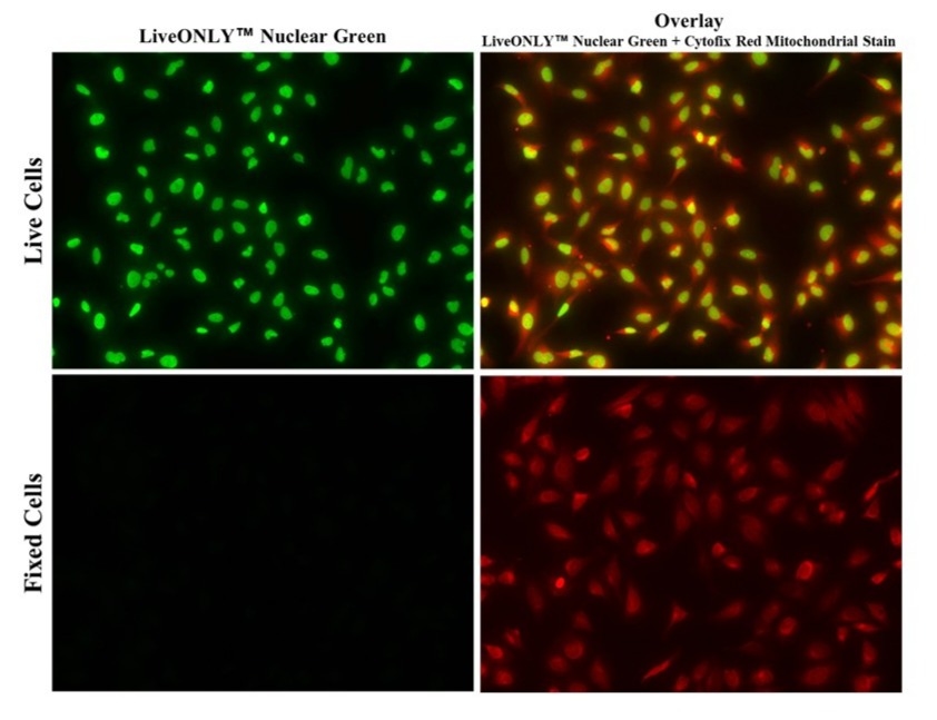
LiveONLY™ Nuclear Green
LiveONLY™ Nuclear Green™ is a cell-permeable nuclear stain that exclusively labels live cell nuclei while excluding dead or membrane-compromised cells, enabling accurate viability assessment in heterogeneous populations.
- First-in-class: First fluorescent dye designed for nuclear staining exclusively in live cells.
- Selective Live Nuclear Staining: Specifically stains nuclei in live cells without background signal from cytoplasm or dead cells
- Live Cell Compatible: Membrane-permeable allowing for real-time imaging of live cells
- No Wash Formula: Simple mix-and-read protocol, eliminates the need for washes
- Bright and Versatile: Bright green signal, compatible with a variety of imaging systems
| Catalog | Size | Price | Quantity |
|---|---|---|---|
| 17687 | 200 tests | Price |

Physical properties
| Solvent | DMSO |
Spectral properties
| Excitation (nm) | 503 |
| Emission (nm) | 527 |
Storage, safety and handling
| H-phrase | H303, H313, H333 |
| Hazard symbol | XN |
| Intended use | Research Use Only (RUO) |
| R-phrase | R20, R21, R22 |
| Storage | Freeze (< -15 °C); Minimize light exposure |
Instrument settings
| Fluorescence microscope | |
| Excitation | FITC filter |
| Emission | FITC filter |
| Recommended plate | Black wall/clear bottom |
Contact us
| Telephone | |
| Fax | |
| sales@aatbio.com | |
| International | See distributors |
| Bulk request | Inquire |
| Custom size | Inquire |
| Technical Support | Contact us |
| Request quotation | Request |
| Purchase order | Send to sales@aatbio.com |
| Shipping | Standard overnight for United States, inquire for international |
Page updated on July 6, 2026