logo
AAT Bioquest

LysoBrite™ Green

Lysosomes are cellular organelles which contain acid hydrolase enzymes to break up waste materials and cellular debris. Lysosomes digest excess or worn-out organelles, food particles, and engulfed viruses or bacteria. The membrane around a lysosome allows the digestive enzymes to work at pH 4.5. The interior of the lysosomes is acidic (pH 4.5-4.8) compared to the slightly alkaline cytosol (pH 7.2). The lysosome maintains this pH differential by pumping protons from the cytosol across the membrane via proton pumps and chloride ion channels. LysoBrite™ Green selectively accumulates in lysosomes probably via the lysosome pH gradient. The lysotropic indicator is a hydrophobic compound that easily permeates intact live cells, and trapped in lysosomes after it gets into cells. Its fluorescence is significantly enhanced upon entering lysosomes. This key feature significantly reduces its staining background and makes it useful for a variety of studies, including cell adhesion, chemotaxis, multidrug resistance, cell viability, apoptosis and cytotoxicity. It is suitable for proliferating and non-proliferating cells, and can be used for both suspension and adherent cells.
Example protocol

AT A GLANCE

Assay Protocol with LysoBrite™ Green
  1. Prepare cells.

  2. Add dye working solution.

  3. Incubate at 37 °C for 30 minutes.

  4. Wash the cells.

  5. Analyze under a fluorescence microscope.

Storage and Handling Conditions

The LysoBrite™ Green stock solution provided is 500X in DMSO. It should be stable for at least 6 months if stored at -20°C. Protect from light, and avoid freeze/thaw cycles.  

PREPARATION OF WORKING SOLUTION

Prepare LysoBrite™ Green Working Solution
  1. Warm LysoBrite™ Green dye to room temperature.

  2. Prepare dye working solution by diluting 20 µL of 500X LysoBrite™ Green with 10 mL of Hanks and 20 mM HEPES buffer (HBSS) or buffer of your choice.

    Note: 20 µL of LysoBrite™ Green dye is enough for one 96-well plate. Aliquot and store unused LysoBrite™ dye stock solutions at < -15 °C. Protect it from light and avoid repeated freeze-thaw cycles.

    Note: The optimal concentration of the fluorescent lysosome indicator varies depending on the specific application. The staining conditions may be modified according to the particular cell type and the permeability of the cells or tissues to the probe. 

SAMPLE EXPERIMENTAL PROTOCOL

This protocol only provides a guideline and should be modified according to your specific needs.

Protocol for Preparing and Staining Adherent Cells
  1. Grow cells in a 96-well black wall/clear bottom plate (100 µL/well/96-well plate) or on coverslips inside a petri dish filled with the appropriate culture medium.

  2. When cells reach the desired confluence, add an equal volume of the dye-working solution (from Preparation of Working Solution Step 2). 

  3. Incubate the cells in a 37 °C, 5% CO2 incubator for 30 minutes.

  4. Wash the cells twice with pre-warmed (37 °C) Hanks and 20 mM HEPES buffer (HBSS) or buffer of your choice. Then fill the cell wells with HBSS or growth medium.

  5. Observe the cells using a fluorescence microscope fitted with the desired filter set.

    Note: It is recommended to increase either the labeling concentration or the incubation time to allow the dye to accumulate if the cells do not appear to be sufficiently stained.

Protocol for Preparing and Staining Suspension Cells
  1. Add an equal volume of the dye-working solution (from Preparation of Working Solution Step 2). 

  2. Incubate the cells in a 37 °C, 5% CO2 incubator for 30 minutes.

  3. Wash the cells twice with pre-warmed (37 °C) Hanks and 20 mM HEPES buffer (HBSS) or buffer of your choice. Then fill the cell wells with HBSS or growth medium.

  4. Observe the cells using a fluorescence microscope fitted with the desired filter set.

    Note: It is recommended to increase either the labeling concentration or the incubation time to allow the dye to accumulate if the cells do not appear to be sufficiently stained.

    Note: Suspension cells may be attached to coverslips treated with BD Cell-Tak® (BD Biosciences) and stained as adherent cells (see Protocol for Preparing and Staining Adherent Cells).

Spectrum

Product family

NameExcitation (nm)Emission (nm)
LysoBrite™ Blue434480
LysoBrite™ Orange543565
LysoBrite™ Red576596
LysoBrite™ Deep Red597619
LysoBrite™ NIR636651

Citations

View all 10 citations: Citation Explorer
mRNA lipid nanoparticle-mediated pyroptosis sensitizes immunologically cold tumors to checkpoint immunotherapy
Authors: Li, Fengqiao and Zhang, Xue-Qing and Ho, William and Tang, Maoping and Li, Zhongyu and Bu, Lei and Xu, Xiaoyang
Journal: Nature Communications (2023): 4223
Synthesis of Dual-Emitting CdZnSe/Mn: ZnS Quantum Dots for Sensing the pH Change in Live Cells
Authors: Mao, Guobin and Wu, Guoqiang and Chen, Minghai and Yan, Chuang and Tang, Jingya and Ma, Yingxin and Zhang, Xian-En
Journal: Analytical Chemistry (2022): 6665--6671
Two-Pronged Intracellular Co-Delivery of Antigen and Adjuvant for Synergistic Cancer Immunotherapy
Authors: Meng, Junli and Zhang, Peisen and Chen, Qizhe and Wang, Zihua and Gu, Yuan and Ma, Jie and Li, Wang and Yang, Chen and Qiao, Yuanyuan and Hou, Yi and others,
Journal: Advanced Materials (2022): 2202168
HIV-1 Tat endocytosis and retention in endolysosomes affects HIV-1 Tat-induced LTR transactivation in astrocytes
Authors: Khan, Nabab and Halcrow, Peter W and Afghah, Zahra and Baral, Aparajita and Geiger, Jonathan D and Chen, Xuesong
Journal: The FASEB Journal (2022): e22184
Microencapsulated Multi-functionalized Graphene Oxide Equipped With Chloroquine for Efficient and Sustained Sirna Delivery
Authors: Imani, Rana and Prakash, Satya and Vali, Hojatollah and Presley, John F and Faghihi, Shahab
Journal: (2021)

References

View all 26 references: Citation Explorer
Requirements, features, and performance of high content screening platforms
Authors: Gough AH, Johnston PA.
Journal: Methods Mol Biol (2007): 41
A pharmaceutical company user's perspective on the potential of high content screening in drug discovery
Authors: Hoffman AF, Garippa RJ.
Journal: Methods Mol Biol (2007): 19
Optimizing the integration of immunoreagents and fluorescent probes for multiplexed high content screening assays
Authors: Giuliano KA., undefined
Journal: Methods Mol Biol (2007): 189
Past, present, and future of high content screening and the field of cellomics
Authors: Taylor DL., undefined
Journal: Methods Mol Biol (2007): 3
Evaluation of a high-content screening fluorescence-based assay analyzing the pharmacological modulation of lipid homeostasis in human macrophages
Authors: Werner T, Liebisch G, Gr and l M, Schmitz G.
Journal: Cytometry A (2006): 200
Page updated on October 6, 2024

Ordering information

Price
Unit size
Catalog Number22643
Quantity
Add to cart

Additional ordering information

Telephone1-800-990-8053
Fax1-800-609-2943
Emailsales@aatbio.com
InternationalSee distributors
Bulk requestInquire
Custom sizeInquire
Technical SupportContact us
Purchase orderSend to sales@aatbio.com
ShippingStandard overnight for United States, inquire for international
Request quotation

Physical properties

Molecular weight

453.34

Solvent

DMSO

Spectral properties

Excitation (nm)

501

Emission (nm)

510

Storage, safety and handling

Certificate of OriginDownload PDF
H-phraseH303, H313, H333
Hazard symbolXN
Intended useResearch Use Only (RUO)
R-phraseR20, R21, R22

Storage

Freeze (< -15 °C); Minimize light exposure
UNSPSC12352200

Platform

Flow cytometer

Excitation488 nm laser
Emission530, 30 nm filter
Instrument specification(s)FITC channel

Fluorescence microscope

ExcitationFITC filter set
EmissionFITC filter set
Recommended plateBlack wall, clear bottom
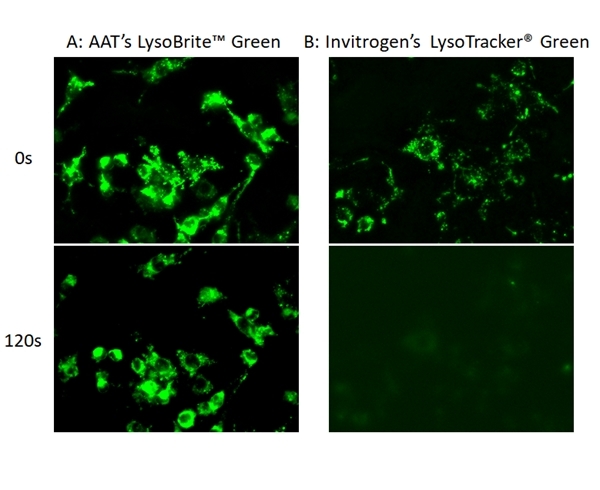
Images of HeLa cells stained with A: AAT&rsquo;s LysoBrite &trade; Green, B: Invitrogen&rsquo;s LysoTracker&reg; Green DND-26 in a Costar black wall/clear bottom 96-well plate. Samples were continuously illuminated for 120 seconds, and the signals were compared before and after the exposure by using a Keyence fluorescence microscope.