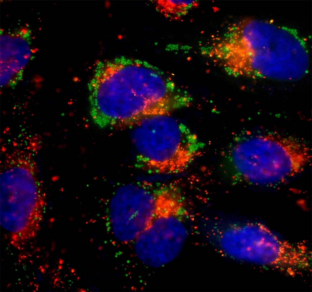
LysoBrite™ Orange
Price | |
Catalog Number | |
Unit Size | |
Quantity |
Telephone | 1-800-990-8053 |
Fax | 1-800-609-2943 |
sales@aatbio.com | |
International | See distributors |
Bulk request | Inquire |
Custom size | Inquire |
Shipping | Standard overnight for United States, inquire for international |
Molecular weight | 670.89 |
Solvent | DMSO |
Excitation (nm) | 543 |
Emission (nm) | 565 |
H-phrase | H303, H313, H333 |
Hazard symbol | XN |
Intended use | Research Use Only (RUO) |
R-phrase | R20, R21, R22 |
Storage | Freeze (< -15 °C); Minimize light exposure |
UNSPSC | 12352200 |
LysoBrite™ Red DND-99 |
LysoBrite™ Green DND-26 |
Overview | ![]() ![]() |
Molecular weight 670.89 | Excitation (nm) 543 | Emission (nm) 565 |
Platform
Flow cytometer
Excitation | 532 nm laser |
Emission | 575/26 nm filter |
Instrument specification(s) | PE channel |
Fluorescence microscope
Excitation | TRITC filter set |
Emission | TRITC filter set |
Recommended plate | Black wall/clear bottom |
Fluorescence microplate reader
Excitation | 520 |
Emission | 550 |
Cutoff | 530 |
Recommended plate | Black wall/clear bottom |
Instrument specification(s) | Bottom read mode |
Example protocol
AT A GLANCE
- Prepare cells
Add dye working solution
Incubate at 37 °C for 30 minutes
- Wash the cells
Analyze under a fluorescence microscope
The LysoBrite™ Orange stock solution provided is 500X in DMSO. It should be stable for at least 6 months if stored at -20°C. Protect from light, and avoid freeze/thaw cycles.
PREPARATION OF WORKING SOLUTION
Warm LysoBrite™ Orange dye to room temperature.
Prepare dye working solution by diluting 20 µL of 500X LysoBrite™ Orange with 10 mL of Hanks and 20 mM HEPES buffer (HBSS) or buffer of your choice.
Note: 20 µL of LysoBrite™ Orange dye is enough for one 96-well plate. Aliquot and store unused LysoBrite™ dye stock solutions at < -15 °C. Protect it from light and avoid repeated freeze-thaw cycles.
Note: The optimal concentration of the fluorescent lysosome indicator varies depending on the specific application. The staining conditions may be modified according to the particular cell type and the permeability of the cells or tissues to the probe.
SAMPLE EXPERIMENTAL PROTOCOL
This protocol only provides a guideline and should be modified according to your specific needs.
Grow cells in a 96-well black wall/clear bottom plate (100 µL/well/96-well plate) or on coverslips inside a petri dish filled with the appropriate culture medium.
When cells reach the desired confluence, add an equal volume of the dye-working solution (from Preparation of Working Solution Step 2).
Incubate the cells in a 37 °C, 5% CO2 incubator for 30 minutes.
Wash the cells twice with pre-warmed (37 °C) Hanks and 20 mM HEPES buffer (HBSS) or buffer of your choice. Then fill the cell wells with HBSS or growth medium.
Observe the cells using a fluorescence microscope fitted with the desired filter set.
Note: It is recommended to increase either the labeling concentration or the incubation time to allow the dye to accumulate if the cells do not appear to be sufficiently stained.
Add an equal volume of the dye-working solution (from Preparation of Working Solution Step 2).
Incubate the cells in a 37 °C, 5% CO2 incubator for 30 minutes.
Wash the cells twice with pre-warmed (37 °C) Hanks and 20 mM HEPES buffer (HBSS) or buffer of your choice. Then fill the cell wells with HBSS or growth medium.
Observe the cells using a fluorescence microscope fitted with the desired filter set.
Note: It is recommended to increase either the labeling concentration or the incubation time to allow the dye to accumulate if the cells do not appear to be sufficiently stained.
Note: Suspension cells may be attached to coverslips treated with BD Cell-Tak® (BD Biosciences) and stained as adherent cells (see Protocol for Preparing and Staining Adherent Cells).
Calculators
Common stock solution preparation
0.1 mg | 0.5 mg | 1 mg | 5 mg | 10 mg | |
1 mM | 149.056 µL | 745.279 µL | 1.491 mL | 7.453 mL | 14.906 mL |
5 mM | 29.811 µL | 149.056 µL | 298.111 µL | 1.491 mL | 2.981 mL |
10 mM | 14.906 µL | 74.528 µL | 149.056 µL | 745.279 µL | 1.491 mL |
Molarity calculator
Mass (Calculate) | Molecular weight | Volume (Calculate) | Concentration (Calculate) | Moles | ||||
/ | = | x | = |
Product Family
Name | Excitation (nm) | Emission (nm) |
LysoBrite™ Blue | 434 | 480 |
LysoBrite™ Green | 501 | 510 |
LysoBrite™ Red | 576 | 596 |
LysoBrite™ Deep Red | 597 | 619 |
LysoBrite™ NIR | 636 | 651 |
Images

Citations
Authors: Thiel, Cora S and ra , undefined and Tauber, Svantje and Lauber, Beatrice and Polzer, Jennifer and Seebacher, Christian and Uhl, Rainer and Neelam, Srujana and Zhang, Ye and Levine, Howard and Ullrich, Oliver
Journal: International journal of molecular sciences (2019): 2402
Authors: Thiel, Cora Sandra and Tauber, Svantje and Lauber, Beatrice and Polzer, Jennifer and Seebacher, Christian and Uhl, Rainer and Neelam, Srujana and Zhang, Ye and Levine, Howard and Ullrich, Oliver
Journal: International journal of molecular sciences (2019): 2402
Authors: Cemma, Marija and Grinstein, Sergio and Brumell, John H
Journal: Autophagy (2016): 1440--1446
Authors: Myung, Ja Hye and Gajjar, Khyati A and Chen, Jihua and Molokie, Robert E and Hong, Seungpyo
Journal: Analytical chemistry (2014): 6088--6094
Authors: Lei, Bo and Chen, Xiaofeng and Han, Xue and Zhou, Jiaan
Journal: Journal of Materials Chemistry (2012): 16906--16913
References
Authors: Gough AH, Johnston PA.
Journal: Methods Mol Biol (2007): 41
Authors: Hoffman AF, Garippa RJ.
Journal: Methods Mol Biol (2007): 19
Authors: Giuliano KA., undefined
Journal: Methods Mol Biol (2007): 189
Authors: Taylor DL., undefined
Journal: Methods Mol Biol (2007): 3
Authors: Martinez ED, Dull AB, Beutler JA, Hager GL.
Journal: Methods Enzymol (2006): 21
Authors: Bowen WP, Wylie PG.
Journal: Assay Drug Dev Technol (2006): 209
Authors: Hudson CC, Oakley RH, Sjaastad MD, Loomis CR.
Journal: Methods Enzymol (2006): 63
Authors: Werner T, Liebisch G, Gr and l M, Schmitz G.
Journal: Cytometry A (2006): 200
Authors: Wolff M, Haasen D, Merk S, Kroner M, Maier U, Bordel S, Wiedenmann J, Nienhaus GU, Valler M, Heilker R.
Journal: Comb Chem High Throughput Screen (2006): 339
Authors: O'Brien P J, Irwin W, Diaz D, Howard-Cofield E, Krejsa CM, Slaughter MR, Gao B, Kaludercic N, Angeline A, Bernardi P, Brain P, Hougham C.
Journal: Arch Toxicol (2006): 580
Application notes
FAQ
Are Cell Navigator® Cell Plasma Membrane Staining Kits suitable for cell culture medium samples?
Are there any alternatives for ethidium bromide in agarose gels?
Are there any alternatives to Cy5?
Are there safer alternatives to ethidium bromide?