Products
Services
Resources
Selection Guides
About
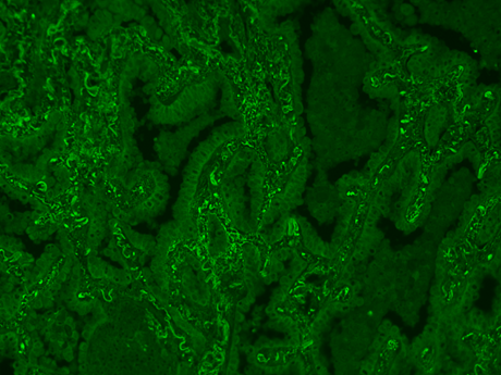
mFluor™ Red 780 Styramide
The Power Styramide™ Signal Amplification (PSA™) system is a highly sensitive method for detecting low-abundant targets in fluorescent immunocytochemistry (ICC), immunohistochemistry (IHC), and in situ hybridization (ISH). By utilizing bright and photostable mFluor™ dyes, Styramide™ conjugates deliver results of unparalleled sharpness and precision, surpassing the sensitivity of standard ICC/IHC/ISH methods by over 100 times, all the while reducing the consumption of primary antibodies. Like tyramide signal amplification (TSA), PSA™ leverages the catalytic activity of horseradish peroxidase (HRP) to generate high-density labeling of a target protein or nucleic acid sequence in situ. The enhanced reactivity of Styramide™ radicals over tyramide ensures faster, more robust labeling of the target, leading to fluorescence signals that are 10-50 times greater than those generated by tyramide (TSA) reagents. The mFluor™ Red 780 Styramide uses the bright red fluorescent dye mFluor™ Red 780 (Ex/Em = 629/767 nm) to label targets in situ. mFluor™ Red 633 is well-excited by the 633 nm laser line and exhibits minimal crosstalk in complex multicolor analysis with blue and green fluorescent probes or other spectrally compatible Styramide conjugates and PSA™ Imaging Kits.
| Catalog | Size | Price | Quantity |
|---|---|---|---|
| 45076 | 100 Slides | Price |

Physical properties
| Solvent | DMSO |
Spectral properties
| Absorbance (nm) | 630 |
| Correction factor (260 nm) | 0.101 |
| Correction factor (280 nm) | 0.116 |
| Extinction coefficient (cm -1 M -1) | 90000 1 |
| Excitation (nm) | 629 |
| Emission (nm) | 767 |
Storage, safety and handling
| H-phrase | H303, H313, H333 |
| Hazard symbol | XN |
| Intended use | Research Use Only (RUO) |
| R-phrase | R20, R21, R22 |
| Storage | Freeze (< -15 °C); Minimize light exposure |
| UNSPSC | 12171501 |
Instrument settings
| Fluorescence microscope | |
| Excitation | 629 nm |
| Emission | 767 nm |
| Recommended plate | Black wall/clear bottom |
Contact us
| Telephone | |
| Fax | |
| sales@aatbio.com | |
| International | See distributors |
| Bulk request | Inquire |
| Custom size | Inquire |
| Technical Support | Contact us |
| Request quotation | Request |
| Purchase order | Send to sales@aatbio.com |
| Shipping | Standard overnight for United States, inquire for international |
Page updated on July 1, 2026