Products
Services
Resources
Selection Guides
About
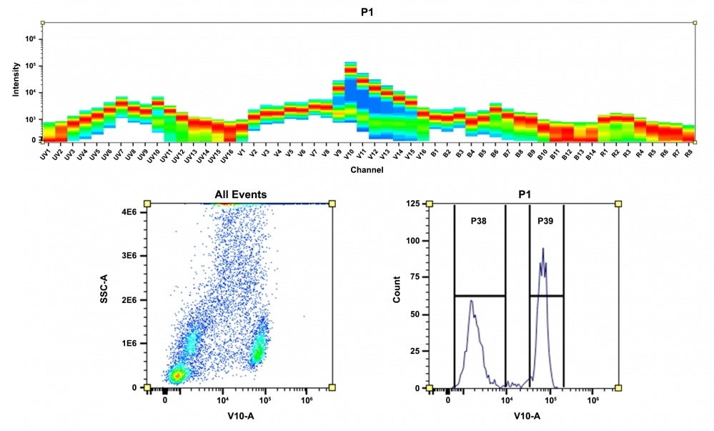
mFluor™ Violet 610 SE
Advances in spectral flow cytometers have expanded applications and capabilities beyond conventional flow cytometry. Now with spectral flow cytometry analysis, researchers and scientists can investigate an increasing number of molecules of interest. However, the potential of spectral flow cytometry is severely limited by the availability of fluorescent labels and readouts. AAT Bioquest's mFluor™ dyes are developed for multicolor flow cytometry-focused applications, in particular, for spectral fluorescence flow cytometry. These dyes have large Stokes Shifts, and can be well excited by the laser lines of flow cytometers (e.g., 350 nm, 405 nm, 488 nm and 633 nm). mFluor™ Violet 610 dyes are well excited with the Violet laser with a red emission ~610 nm. These spectral characteristics make them very unique labeling dyes for spectral fluorescence flow cytometric applications. mFluor™ Violet 610 SE is reasonably stable and shows good reactivity and selectivity with protein amino groups. mFluor™ Violet 610 SE provides a convenient tool to label monoclonal, polyclonal antibodies or other proteins (>10 kDa) for flow cytometric applications with the violet laser excitation.
| Catalog | Size | Price | Quantity |
|---|---|---|---|
| 1156 | 1 mg | Price |

Physical properties
| Molecular weight | 1293.46 |
| Solvent | DMSO |
Spectral properties
| Absorbance (nm) | 594 |
| Correction factor (260 nm) | 0.532 |
| Correction factor (280 nm) | 0.66 |
| Extinction coefficient (cm -1 M -1) | 90000 1 |
| Excitation (nm) | 421 |
| Emission (nm) | 612 |
| Quantum yield | 0.3 1 |
Storage, safety and handling
| H-phrase | H303, H313, H333 |
| Hazard symbol | XN |
| Intended use | Research Use Only (RUO) |
| R-phrase | R20, R21, R22 |
| Storage | Freeze (< -15 °C); Minimize light exposure |
| UNSPSC | 12171501 |
Contact us
| Telephone | |
| Fax | |
| sales@aatbio.com | |
| International | See distributors |
| Bulk request | Inquire |
| Custom size | Inquire |
| Technical Support | Contact us |
| Request quotation | Request |
| Purchase order | Send to sales@aatbio.com |
| Shipping | Standard overnight for United States, inquire for international |
Page updated on July 6, 2026