Products
Services
Resources
Selection Guides
About
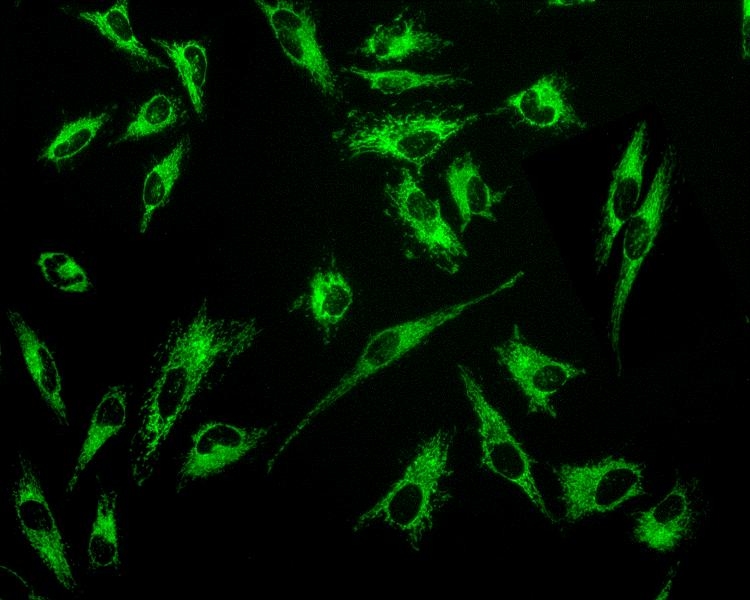
MitoLite™ Green EX488
MitoLite™ Green is a cationic dye that selectively accumulates in mitochondria probably vial the mitochondrial membrane potential gradient. The mitochondrial indicator is a hydrophobic compound that easily permeates intact live cells, and trapped in mitochondria after it gets into cells. This fluorescent mitochondrial indicator is retained in mitochondria for long time since the indicator carries a cell-retaining group. This key feature significantly increases its staining efficiency. The labeling protocol is robust, requiring minimal hands-on time. It can be readily adapted for a wide variety of fluorescence platforms such as microplate assays, immunocytochemistry and flow cytometry. It is suitable for proliferating and non-proliferating cells, and can be used for both suspension and adherent cells.
| Catalog | Size | Price | Quantity |
|---|---|---|---|
| 22675 | 500 Tests | Price |

Physical properties
| Molecular weight | N/A |
| Solvent | DMSO |
Spectral properties
| Excitation (nm) | 508 |
| Emission (nm) | 528 |
Usage and storage
| Intended use | Research Use Only (RUO) |
| Storage | Freeze (< -15 °C); Minimize light exposure |
Contact us
| Telephone | |
| Fax | |
| sales@aatbio.com | |
| International | See distributors |
| Bulk request | Inquire |
| Custom size | Inquire |
| Technical Support | Contact us |
| Request quotation | Request |
| Purchase order | Send to sales@aatbio.com |
| Shipping | Standard overnight for United States, inquire for international |
Page updated on July 16, 2026