logo
AAT Bioquest

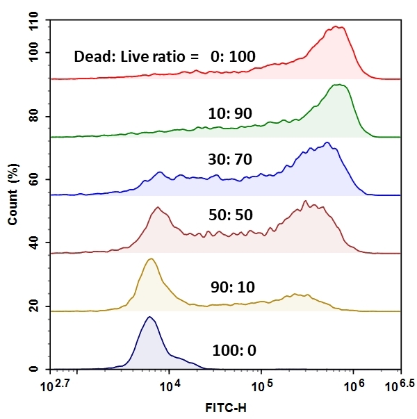
MycoLight™ Flow Cytometric Live Bacteria Assay Kit

Relative viability of <em>E.coli</em> suspension was analyzed using ACEA NovoCyte flow cytometer in FITC channel. The readings (Count(%)) were obtained from various Live/Dead <em>E.coli</em> mixtures (Table 1). The live and dead population in each mixture can be easily distinguished by the two distinct peaks.
Ordering information
Price
Catalog Number
Unit Size
Quantity
Add to cart
Additional ordering information
Telephone1-800-990-8053
Fax1-800-609-2943
Emailsales@aatbio.com
InternationalSee distributors
Bulk requestInquire
Custom sizeInquire
ShippingStandard overnight for United States, inquire for international
Request quotation
Spectral properties
Correction Factor (260 nm)0.31
Correction Factor (280 nm)0.12
Extinction coefficient (cm -1 M -1)76000
Excitation (nm)498
Emission (nm)526
Storage, safety and handling
H-phraseH303, H313, H333
Hazard symbolXN
Intended useResearch Use Only (RUO)
R-phraseR20, R21, R22
UNSPSC12352200

OverviewpdfSDSpdfProtocol


Correction Factor (260 nm)
0.31
Correction Factor (280 nm)
0.12
Extinction coefficient (cm -1 M -1)
76000
Excitation (nm)
498
Emission (nm)
526
The MycoLight Flow Cytometric Live Bacteria Assay Kit provides an easy and convenient methodfor evaluating bacterial vitality as a function of the intracellular esterase activity. MycoLight™ 520 is non-fluorescent esterase substrate that diffuse into both Gram positive and Gram-negative bacteria. Upon hydrolysis by bacterial intracellular non-specific esterase, a green fluorescent product is produced and accumulated within bacteria. Compared to the commonly used esterase substrate CFDA and CFDA-AM, this kit provides brighter and more stable signal with lower background and easier staining protocol.

Platform


Flow cytometer

Excitation488 nm laser
Emission530/30 nm filter
Instrument specification(s)FITC channel

Components


Example protocol


AT A GLANCE

Protocol summary

  1. Prepare 100X dye stock solution.
  2. Prepare bacteria samples.
  3. Incubate bacteria samples with MycoLight™ 520 at 37°C for 5-10 minutes or room temperature for 60 minutes in dark.
  4. Analyze sample by flow cytometry with FITC channel.

Important
Thaw one of each kit component at room temperature before starting the experiment.

PREPARATION OF STOCK SOLUTION

Unless otherwise noted, all unused stock solutions should be divided into single-use aliquots and stored at -20 °C after preparation. Avoid repeated freeze-thaw cycles.

1. MycoLight™ 520 stock solution (100X):
Add 100 µL of DMSO (Component C) into one vial of MycoLight™ 520 (Component A) to make 100X stock solution.


SAMPLE EXPERIMENTAL PROTOCOL

  1. Prepare bacteria sample with concentration in range of 106 to 108 cells/ml. Grow bacteria into late log phase in appropriate medium. Remove medium by centrifugation at 10,000 x g for 10 minutes and re-suspend the pellet in Assay Buffer (Component B).  Note: Measure the optical density of the bacterial culture at wavelength = 600 nm (OD600) to determine the cell number. For E. coli culture, OD600 = 1.0 equals 8 x 10⁸ cells/ml.                                                                                                                                                                                                  
  2. Treat cells with test compounds as desired. Remove treatments by centrifugation at 10,000 x g for 10 minutes and re-suspend the pellet in appropriate amount of Assay Buffer (Component B) so the concentration of bacteria in the treated sample is the same as the live.  Note:  Determine the concentration of the bacterial culture before starting the treatment. Note: Dead bacteria can serve as negative control, it is recommended to kill bacteria with 70% ethanol for 30 min followed by 60 min of boiling. 
                                                                                                                                                                                                                                       
  3. Example of Live/Dead bacterial mixtrue preparation: Mix seven different propotions of the bacterial suspensions as in Table 1 for a total volume of 200 µL for each sample.                                                                                                                                                                                                                                                   
  4. Add 2 µL of the 100X MycoLight™ 520 stock solution to 200 µL of the bacterial sample in Assay Buffer.                                                                                                                                                                                                              
  5. Mix well and incubate in dark for 5-10 min at 37°C or 60 min at RT for optimum staining results.                                                                                                                                                                                                   
  6. Add 300 µL of Assay Buffer (Component C) to increase volume before analyzing the cells with a flow cytometer.                                                                                                                                                                       
  7. Monitor fluorescence of bacteria with a flow cytometer through FITC channel (488/530 nm).  Note: To exclude debris, it is recommended to set the threshold of the flow cytometer as the following: FSC >10,000, SSC>5,000. Note: The efficiency of MycoLight™ 520 is highly strain dependent and the staining conditions should be optimized accordingly.


Table 1. Example Volumes of Live and Dead samples to mix to achieve various Live/Dead ratios.

Ratio of Dead:Live CellsµL of Dead SampleµL of Live Sample
0:1000200
10:9020180
30:7060140
50:50100100
90:1018020
100:02000


Spectrum


Open in Advanced Spectrum Viewer
spectrum

Spectral properties

Correction Factor (260 nm)0.31
Correction Factor (280 nm)0.12
Extinction coefficient (cm -1 M -1)76000
Excitation (nm)498
Emission (nm)526

Images


Citations


View all 13 citations: Citation Explorer
Seminal plasma affects the survival rate and motility pattern of raw llama spermatozoa
Authors: Fumuso, F. G., Giuliano, S. M., Chaves, M. G., Neild, D. M., Miragaya, M. H., Gambarotta, M. C., Carretero, M. I.
Journal: Anim Reprod Sci (2018): 99-106
New fluorescein precursors for live bacteria detection
Authors: Guilini, C., Baehr, C., Schaeffer, E., Gizzi, P., Rufi, F., Haiech, J., Weiss, E., Bonnet, D., Galzi, J. L.
Journal: Anal Chem (2015): 8858-66
Protective effects of antithrombin on free groin flaps after secondary venous stasis in the rat model
Authors: Wallmichrath, J., Knab, R., Baumeister, R. G., Volkmer, E., Giunta, R. E., Frick, A.
Journal: J Plast Reconstr Aesthet Surg (2014): 707-11
Comparison of different live/dead stainings for detection and quantification of adherent microorganisms in the initial oral biofilm
Authors: Tawakoli, P. N., Al-Ahmad, A., Hoth-Hannig, W., Hannig, M., Hannig, C.
Journal: Clin Oral Investig (2013): 841-50
A multiplexed immunofluorescence method identifies Phakopsora pachyrhizi Urediniospores and determines their viability
Authors: Vittal, R., Haudenshield, J. S., Hartman, G. L.
Journal: Phytopathology (2012): 1143-52
Rapid flow cytometric method for viability determination of solventogenic clostridia
Authors: Linhova, M., Branska, B., Patakova, P., Lipovsky, J., Fribert, P., Rychtera, M., Melzoch, K.
Journal: Folia Microbiol (Praha) (2012): 307-11
Expansion of chondrocytes in a three-dimensional matrix for tracheal tissue engineering
Authors: Walles, T., Giere, B., Macchiarini, P., Mertsching, H.
Journal: Ann Thorac Surg (2004): 444-8; discussion 448-9
Functional changes and motility characteristics of Japanese Black bull spermatozoa separated by Percoll
Authors: Suzuki, K., Geshi, M., Yamauchi, N., Nagai, T.
Journal: Anim Reprod Sci (2003): 157-72
Validation of flow cytometry for assessment of viability and acrosomal integrity of dog spermatozoa and for evaluation of different methods of cryopreservation
Authors: Pena, A. I., Johannisson, A., Linde-Forsberg, C.
Journal: J Reprod Fertil Suppl (2001): 371-6
Kinetic and temporal factors influence chilling injury to germinal vesicle and mature bovine oocytes
Authors: Zeron, Y., Pearl, M., Borochov, A., Arav, A.
Journal: Cryobiology (1999): 35-42