logo
AAT Bioquest

MycoLight™ Red JJ94 *2.5 mM in DMSO*

Optical density (OD600 nm) of <em>E. coli</em> LB cultures supplemented with 1% DMSO (control), MycoLight<sup>TM</sup>&nbsp; Red JJ94 or Syto-9<sup>TM</sup> &nbsp;over time. Syto-9<sup>TM</sup> inhibits bacterial growth substantially while MycoLight&trade; Red JJ94 is fully compatible with normal bacterial growth.
<em>Rhodococcus</em> <em>qingshengii</em> was stained with 2.5 &mu;M of MycoLight&trade; Red JJ94 for 20 minutes. Image was taken by Keyence florescent microscope with Cy5 filter set.
Ordering information
Price
Catalog Number
Unit Size
Quantity
Add to cart
Additional ordering information
Telephone1-800-990-8053
Fax1-800-609-2943
Emailsales@aatbio.com
InternationalSee distributors
Bulk requestInquire
Custom sizeInquire
ShippingStandard overnight for United States, inquire for international
Request quotation
Physical properties
Molecular weight738.75
SolventDMSO
Storage, safety and handling
H-phraseH301, H311, H331
Hazard symbolT
Intended useResearch Use Only (RUO)
R-phraseR23, R24, R25
StorageFreeze (< -15 °C); Minimize light exposure
UNSPSC12352200

OverviewpdfSDSpdfProtocol


Molecular weight
738.75
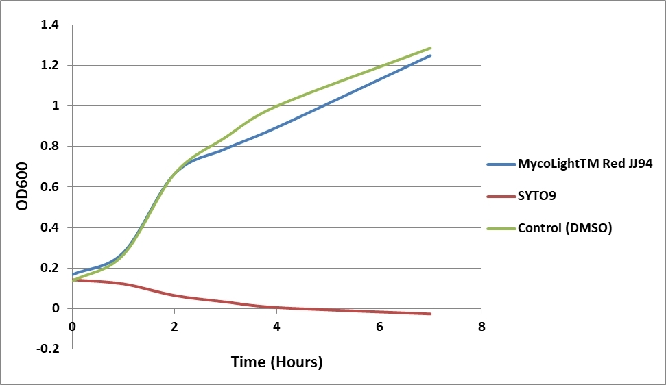
Green fluorescent protein (GFP) derivatives are widely used as fluorescent reporters to study biological processes at the cellular and molecular level, including labeling bacteria. Unfortunately, the GFP-based bacterial labeling is tedious, and several important organisms have remained difficult to modify for fluorescent protein expression. Complimentary to GFP-based fluorescence labeling of bacteria, SYTO-9 has been widely used for labeling bacteria in a more convenient mode, i.e., through a simple incubation. However, SYTO-9 is reported to significantly inhibit some bacterial growth. AAT Bioquest has developed MycoLight™ Red JJ94 that has minimal inhibition of bacterial growth compared to SYTO-9. In addition, MycoLight™ Red JJ94 can be used for multicolor detection of bacteria with the widely used GFP probes. It has an excitation and emission at ~637 and ~651 nm, respectively. This perfectly matches the He-laser excitation and Cy5 filter set that are commonly equipped in most fluorescence microscopes and flow cytometers. MycoLight™ Red JJ94 is a far-red fluorescent DNA-binding dye for straightforward live bacterial cell labeling, allowing subsequent infection studies. It has low intrinsic fluorescence, but upon binding DNA, its fluorescence emission intensity increases dramatically. MycoLight™ Red JJ94 readily stains live bacteria without compromising microbial growth. Under the same conditions, SYTO-9 inhibits bacterial growth substantially while MycoLight™ Red JJ94 is fully compatible with normal bacterial growth in liquid medium or on solid medium. The bacterial fluorescence labeling by MycoLight™ Red JJ94 is stable over several hours after shifting the bacteria into dye-free media.

Platform


Flow cytometer

Excitation640 nm laser
Emission660/20 nm filter
Instrument specification(s)APC channel

Fluorescence microscope

Excitation630 nm
Emission660 nm
Recommended plateBlack wall/clear bottom
Instrument specification(s)Cy5 filterset

Example protocol


AT A GLANCE

  1. Stains live bacteria without affecting normal microbial growth
  2. Uses Cy5 filter set and He-laser excitation, widely available in most flow cytometers and fluorescent microscope
  3. Can be used for multicolor detection in bacteria
  4. Stable labelling for several hours 
Important Thaw the components at room temperature before starting the experiment.

SAMPLE EXPERIMENTAL PROTOCOL

  1. Dilute the MycoLight™ Red JJ94 with buffer of your choice. MycoLight™ Red JJ94 can be supplemented directly into bacterial culture medium to the final concentration of 2.5 µM.
  2. Vortex samples to mix, and then incubate for at least 10 minutes.
  3. Monitor sample florescent signal by florescent microscope or flow cytometer with Cy5 filter set. 

Note      The above protocol can be adapted for most bacterial strains. These conditions require adjustment for each strain and experimental system. Growth medium, cell density, the presence of other organisms and factors may influence staining. Residual detergent on glassware may also affect staining of many organisms, and cause brightly stained material to appear in solutions with or without cells present.
Note      Use plastic tubes when diluting MycoLight™ Red JJ94, because the diluted stain adheres to glass. In general, the best results are obtained in buffers that do not contain phosphate.
Note      Dye concentration may be optimized with different bacterial strain to obtain best results.
Note      Since MycoLight™ Red JJ94 is fully compatible with bacterial growth, live bacteria can be incubated with MycoLight™ Red JJ94 for prolonged period of time.

Calculators


Common stock solution preparation

Table 1. Volume of DMSO needed to reconstitute specific mass of MycoLight™ Red JJ94 *2.5 mM in DMSO* to given concentration. Note that volume is only for preparing stock solution. Refer to sample experimental protocol for appropriate experimental/physiological buffers.

0.1 mg0.5 mg1 mg5 mg10 mg
1 mM135.364 µL676.819 µL1.354 mL6.768 mL13.536 mL
5 mM27.073 µL135.364 µL270.728 µL1.354 mL2.707 mL
10 mM13.536 µL67.682 µL135.364 µL676.819 µL1.354 mL

Molarity calculator

Enter any two values (mass, volume, concentration) to calculate the third.

Mass (Calculate)Molecular weightVolume (Calculate)Concentration (Calculate)Moles
/=x=

Images


References


View all 25 references: Citation Explorer
A loop-mediated isothermal amplification (LAMP) assay for Strongyloides stercoralis in stool that uses a visual detection method with SYTO-82 fluorescent dye
Authors: Watts MR, James G, Sultana Y, Ginn AN, Outhred AC, Kong F, Verweij JJ, Iredell JR, Chen SC, Lee R.
Journal: Am J Trop Med Hyg (2014): 306
A new triplex real time PCR which distinguishes between MRSA, MSSA, and mecA coagulase negative strains by means of melting point analysis using SYTO 9
Authors: Weidner J, Cassens U, Gohde W, Wullenweber J, Greve B.
Journal: Clin Lab (2013): 795
Compatibility of SYTO 13 and Hoechst 33342 for longitudinal imaging of neuron viability and cell death
Authors: Hubbard KS, Gut IM, Scheeler SM, Lyman ME, McNutt PM.
Journal: BMC Res Notes (2012): 437
Evaluation of Pseudomonas aeruginosa (PAO1) adhesion to human alveolar epithelial cells A549 using SYTO 9 dye
Authors: Larrosa M, Truchado P, Espin JC, Tomas-Barberan FA, Allende A, Garcia-Conesa MT.
Journal: Mol Cell Probes (2012): 121
Rapid quantification of cell viability and apoptosis in B-cell lymphoma cultures using cyanine SYTO probes
Authors: Wlodkowic D, Skommer J, Darzynkiewicz Z.
Journal: Methods Mol Biol (2011): 81
SYTO dyes and EvaGreen outperform SYBR Green in real-time PCR
Authors: Eischeid AC., undefined
Journal: BMC Res Notes (2011): 263
Use of SYTO 13, a fluorescent dye binding nucleic acids, for the detection of microparticles in in vitro systems
Authors: Ullal AJ, Pisetsky DS, Reich CF, 3rd.
Journal: Cytometry A (2010): 294
Detection of methicillin-resistant Staphylococcus aureus using double duplex real-time PCR and dye Syto 9
Authors: Seputiene V, Vilkoicaite A, Armalyte J, Pavilonis A, Suziedeliene E.
Journal: Folia Microbiol (Praha) (2010): 502
Validation of SYTO 9/propidium iodide uptake for rapid detection of viable but noncultivable Legionella pneumophila
Authors: Giao MS, Wilks SA, Azevedo NF, Vieira MJ, Keevil CW.
Journal: Microb Ecol (2009): 56
Quantitative measurement of Plasmodium-infected erythrocytes in murine models of malaria by flow cytometry using bidimensional assessment of SYTO-16 fluorescence
Authors: Jimenez-Diaz MB, Mulet T, Gomez V, Viera S, Alvarez A, Garuti H, Vazquez Y, Fern and ez A, Ibanez J, Jimenez M, Gargallo-Viola D, Angulo-Barturen I.
Journal: Cytometry A (2009): 225