logo
AAT Bioquest

Nuclear Blue™ DCS2 *Equivalent to SYTOX™ Blue Dead Cell Stain*

A mixture of heat-killed and untreated Jurkat cells was stained with Nuclear Blue™ DCS2 (17546) or Nuclear Violet™ DCS1 (17549) stain for 10 minutes. Cells were analyzed on a flow cytometer equipped with a 405 nm violet laser and a 473/15 nm bandpass filter, such as the V4 channel on Cytek's Aurora spectral flow cytometer. Live cells are easily distinguished from the dead cell population.
Ordering information
Price
Catalog Number
Unit Size
Quantity
Add to cart
Additional ordering information
Telephone1-800-990-8053
Fax1-800-609-2943
Emailsales@aatbio.com
InternationalSee distributors
Bulk requestInquire
Custom sizeInquire
ShippingStandard overnight for United States, inquire for international
Request quotation
Physical properties
Molecular weight668.36
SolventDMSO
Spectral properties
Excitation (nm)445
Emission (nm)470
Storage, safety and handling
H-phraseH303, H313, H333
Hazard symbolXN
Intended useResearch Use Only (RUO)
R-phraseR20, R21, R22
StorageFreeze (< -15 °C); Minimize light exposure

OverviewpdfSDSpdfProtocol


See also: Nucleus
Molecular weight
668.36
Excitation (nm)
445
Emission (nm)
470
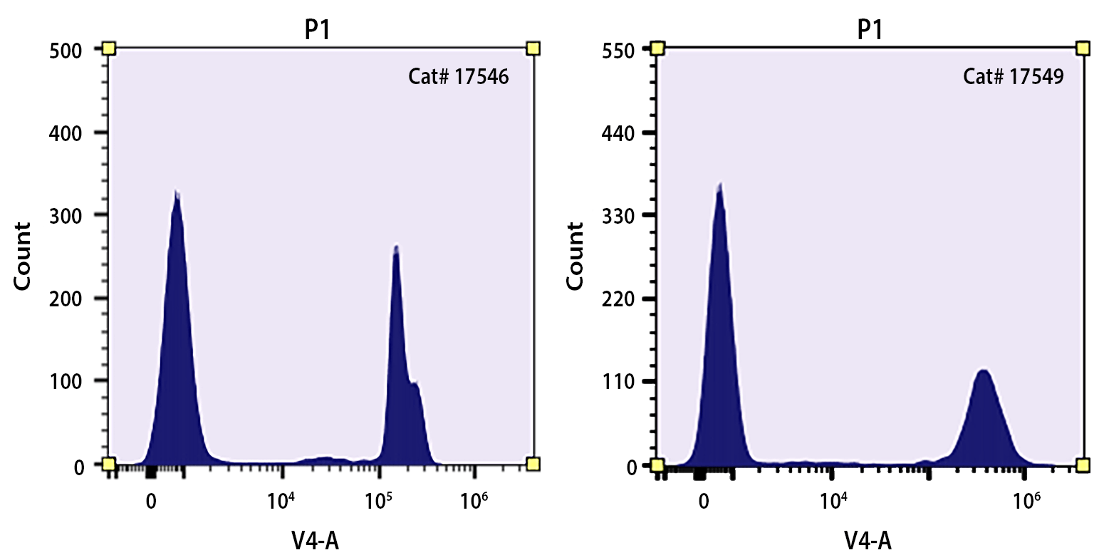
Nuclear Blue™ DCS2 is the same molecule to SYTOX™ Blue Dead Cell Stain (SYTOX™ is a trademark of ThermoFisher). Nuclear Blue™ DCS2 is a simple and quantitative single-step dead-cell indicator for use with violet laser equipped flow cytometers. It is a high-affinity nucleic acid stain that easily penetrates cells with compromised plasma membranes but will not cross uncompromised cell membranes. Under the same conditions, our Nuclear Violet™ DCS1 (#17549) gives much higher signal/background ratio than SYTOX™ Blue Dead Cell Stain. Nuclear Violet™ DCS1 is better excited by the violet laser at 405 nm than SYTOX™ Blue Dead Cell Stain. After brief incubation with Nuclear Violet™ DCS1 stain, the nucleic acids of dead cells fluoresce bright blue when excited with 405 nm violet laser light. The violet-excited fluorescence emission of Nuclear Violet™ DCS1 stain permits clear discrimination from probes excited by most other laser lines, facilitating the development of multicolor assays with minimal spectral overlap between signals.

Platform


Flow cytometer

Excitation405 nm Laser
Emission473/15 nm Filter

Example protocol


SAMPLE EXPERIMENTAL PROTOCOL

The following protocol can be used as a general guideline and is adaptable for any cell type.

Note: Growth medium, cell density, and other factors may influence staining. For optimal staining, try a range of dye concentrations to determine the one that yields the best results.

Note: Before using, thaw the vial of Nuclear Blue™ DCS2 to room temperature 

  1. Harvest sample cells using an appropriate buffer and adjust the cell concentration of the sample to be from 1 x 10⁵ to 5 x 10⁷ cells/mL.

  2. Prepare flow cytometry tube(s) containing 1 mL of cell suspension.

  3. Add 1 µL of Nuclear Blue™ DCS2 to each flow cytometry tube.

  4. Incubate flow cytometry tubes for 5 to 10 minutes at room temperature.

  5. Analyze samples without washing on a flow cytometer with a 473/15 nm emission filter.

Calculators


Common stock solution preparation

Table 1. Volume of DMSO needed to reconstitute specific mass of Nuclear Blue™ DCS2 *Equivalent to SYTOX™ Blue Dead Cell Stain* to given concentration. Note that volume is only for preparing stock solution. Refer to sample experimental protocol for appropriate experimental/physiological buffers.

0.1 mg0.5 mg1 mg5 mg10 mg
1 mM149.62 µL748.1 µL1.496 mL7.481 mL14.962 mL
5 mM29.924 µL149.62 µL299.24 µL1.496 mL2.992 mL
10 mM14.962 µL74.81 µL149.62 µL748.1 µL1.496 mL

Molarity calculator

Enter any two values (mass, volume, concentration) to calculate the third.

Mass (Calculate)Molecular weightVolume (Calculate)Concentration (Calculate)Moles
/=x=

Spectrum


Open in Advanced Spectrum Viewer
spectrum

Spectral properties

Excitation (nm)445
Emission (nm)470

Product Family


NameExcitation (nm)Emission (nm)
Nuclear Blue™ DCS1 *5 mM DMSO Solution*348469
Nuclear Blue™ LCS1353456

Images


References


View all 50 references: Citation Explorer
Characterization of microglia behaviour in healthy and pathological conditions with image analysis tools.
Authors: Martinez, Aleix and Hériché, Jean-Karim and Calvo, Maria and Tischer, Christian and Otxoa-de-Amezaga, Amaia and Pedragosa, Jordi and Bosch, Anna and Planas, Anna M and Petegnief, Valérie
Journal: Open biology (2023): 220200
A comprehensive method to study the DNA's association with lamin and chromatin compaction in intact cell nuclei at super resolution.
Authors: Chapman, Katarina B and Filipsky, Filip and Peschke, Nicolas and Gelléri, Márton and Weinhardt, Venera and Braun, Andrejs and Hausmann, Michael and Cremer, Christoph
Journal: Nanoscale (2023): 742-756
6-C-Linked trehalose glycolipids signal through Mincle and exhibit potent adjuvant activity.
Authors: Thathsaranie P Manthrirathna, M A and Kodar, Kristel and Ishizuka, Shigenari and Dangerfield, Emma M and Xiuyuan, Lu and Yamasaki, Sho and Stocker, Bridget L and S M Timmer, Mattie
Journal: Bioorganic chemistry (2023): 106345
Asebogenin suppresses thrombus formation via inhibition of Syk phosphorylation.
Authors: Li, Li and Xu, Xulin and Lv, Keyu and Zheng, Guijuan and Wang, Hao and Chen, Shuai and Huang, Lang and Liu, Yi and Zhang, Yadong and Tang, Zhaoming and Zhang, Lili and Wang, Jinyu and Qiao, Jianlin and Li, Hongliang and Wang, Xuanbin and Yao, Guangmin and Fang, Chao
Journal: British journal of pharmacology (2023): 287-307
Effects of Imatinib Combined With Everolimus on Mouse Pituitary Tumor Cell AtT-20.
Authors: Wang, Wenxin and Lin, Yi and Qu, Xinguo and Meng, Linghu and Yang, Jianquan
Journal: Alternative therapies in health and medicine (2023): 238-244
A new fluorescence labeling method for molecular analysis of double-stranded DNA.
Authors: Takahashi, Shunsuke and Oshige, Masahiko and Katsura, Shinji and Nagahara, Yukitoshi
Journal: Analytical biochemistry (2023): 115000
The amoebicidal effect of Torreya nucifera extract on Acanthamoeba lugdunensis.
Authors: Kang, Min Seung and Kim, Sangyoon and Kim, Da Som and Yu, Hak Sun and Lee, Ji Eun
Journal: PloS one (2023): e0281141
Absolute Configuration and Antileishmanial Activity of (-)-Cyclocolorenone Isolated from Duguetia lanceolata (Annonaceae).
Authors: Monteiro, Jackson and Passero, Luiz Felipe D and Jesus, Jéssica A and Laurenti, Márcia D and Lago, João H G and Soares, Marisi G and Batista, Andrea N L and Batista, João M and Sartorelli, Patricia
Journal: Current topics in medicinal chemistry (2022): 1626-1633
Measuring and Perturbing Ferroptosis in Plants.
Authors: Distéfano, Ayelen M and Marchetti, Fernanda and Zabaleta, Eduardo and Pagnussat, Gabriela C
Journal: Methods in molecular biology (Clifton, N.J.) (2022): 185-192
In vitro study of the ecotoxicological risk of methylisothiazolinone and chloroxylenol towards soil bacteria.
Authors: Nowak-Lange, Marta and Niedziałkowska, Katarzyna and Bernat, Przemysław and Lisowska, Katarzyna
Journal: Scientific reports (2022): 19068