logo
AAT Bioquest

PE-iFluor® 700 Tandem

PE-iFluor® 700 Tandem is a new color used in flow cytometry. Its primary absorption peak is at 565 nm with emission peak at ~720 nm. It has been validated with a spectral flow cytometer. PE-iFluor® 700 Tandem gives a significantly improved staining index than the corresponding PE-Alexa Fluor™ 700 Tandem. AAT Bioquest offer the largest number of colors for conventional and spectral flow cytometry applications, including iFluor®, mFluor™ small organic dyes and a variety of their tandems.
Spectrum

Product family

NameExcitation (nm)Emission (nm)Extinction coefficient (cm -1 M -1)
PE-iFluor® 610 Tandem5656251960000
PE-iFluor® 647 Tandem5656661960000
PE-iFluor® 750 Tandem5657781960000
PE-iFluor® 710 Tandem5657471960000
PE-iFluor® 594 Tandem5656061960000
PE-iFluor® 660 Tandem5656951960000
PE-iFluor® 780 Tandem5665751960000
PE-iFluor® 597 Tandem565612-
PE-iFluor® 740 Tandem5657671960000
PE-iFluor® 720 Tandem5657501960000
PE-iFluor® 770 Tandem5675751960000
APC-iFluor® 700 Tandem651710700000
Show More (3)

References

View all 13 references: Citation Explorer
Multifunctional Silica-Based Nanoparticles with Controlled Release of Organotin Metallodrug for Targeted Theranosis of Breast Cancer.
Authors: Ovejero Paredes, Karina and Díaz-García, Diana and García-Almodóvar, Victoria and Lozano Chamizo, Laura and Marciello, Marzia and Díaz-Sánchez, Miguel and Prashar, Sanjiv and Gómez-Ruiz, Santiago and Filice, Marco
Journal: Cancers (2020)
Photobleaching Comparison of R-Phycoerythrin-Streptavidin and Streptavidin-Alexa Fluor 568 in a Breast Cancer Cell Line.
Authors: Ostad, Seyed Nasser and Babaei, Sepideh and Bayat, Ali Ahmad and Mahmoudian, Jafar
Journal: Monoclonal antibodies in immunodiagnosis and immunotherapy (2019): 25-29
[Changes of monocyte and monocyte-platelet aggregates in different subgroups of thrombotic events in patients with acute myocardial infarction during PCI].
Authors: Wang, Sheng and Sun, Cuifang and Liao, Wang and Wu, Zhongwei and Wang, Yudai and Huang, Xiuxian and Lu, Sijia and Dong, Xiaoli and Shuai, Fujie and Li, Bin
Journal: Xi bao yu fen zi mian yi xue za zhi = Chinese journal of cellular and molecular immunology (2017): 959-965
Novel PSCA targeting scFv-fusion proteins for diagnosis and immunotherapy of prostate cancer.
Authors: Kessler, Claudia and Pardo, Alessa and Tur, Mehmet K and Gattenlöhner, Stefan and Fischer, Rainer and Kolberg, Katharina and Barth, Stefan
Journal: Journal of cancer research and clinical oncology (2017): 2025-2038
Combination of CD157 and FLAER to Detect Peripheral Blood Eosinophils by Multiparameter Flow Cytometry.
Authors: Carulli, Giovanni and Marini, Alessandra and Sammuri, Paola and Domenichini, Cristiana and Ottaviano, Virginia and Pacini, Simone and Petrini, Mario
Journal: Journal of clinical and experimental hematopathology : JCEH (2015): 55-60
Page updated on December 11, 2024

Ordering information

Price
Unit size
Catalog Number2614
Quantity
Add to cart

Additional ordering information

Telephone1-800-990-8053
Fax1-800-609-2943
Emailsales@aatbio.com
InternationalSee distributors
Bulk requestInquire
Custom sizeInquire
Technical SupportContact us
Purchase orderSend to sales@aatbio.com
ShippingStandard overnight for United States, inquire for international
Request quotation

Physical properties

Solvent

Water

Spectral properties

Absorbance (nm)

566

Extinction coefficient (cm -1 M -1)

1960000

Excitation (nm)

565

Emission (nm)

708

Storage, safety and handling

H-phraseH303, H313, H333
Hazard symbolXN
Intended useResearch Use Only (RUO)
R-phraseR20, R21, R22

Storage

Refrigerated (2-8 °C); Minimize light exposure
UNSPSC12171501
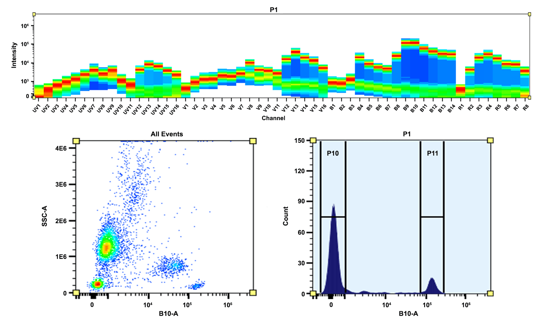
Top) Spectral pattern was generated using a 4-laser spectral cytometer. Spatially offset lasers (355 nm, 405 nm, 488 nm, and 640 nm) were used to create four distinct emission profiles, then, when combined, yielded the overall spectral signature. Bottom) Flow cytometry analysis of PBMC stained with PE/iFlour® 700 anti-human CD4 *SK3* conjugate. The fluorescence signal was monitored using an Aurora spectral flow cytometer in the PE/iFluor® 700 specific B10-A channel.
Flow cytometry analysis of whole blood stained with PE-iFluor® 700 anti-human CD19 *HIB19* conjugate. The fluorescence signal was monitored using an Aurora spectral flow cytometer in the PE-iFluor® 700 specific B10-A channel.