logo
AAT Bioquest

PhosphoWorks™ Colorimetric MESG Phosphate Assay Kit *UV absorption*

In the presence of inorganic phosphate MESG is converted to 2-amino-6-mercapto-7-methlpurine by purine nucleoside phosphorylase (EC 2.4.2.1) with absorption wavelength shift to red. This feature has been used to develop our convenient MESG phosphate assay kit. Our kit provides all the essential reagents including MESG, phosphorylase and reaction buffer. The MESG substrate gives an absorbance increase at 360 nm on phosphorylysis at pH 6.5-8.5, and at pH 7.6 the change in extinction coefficient is 11,000 M-1cm-1. The assay is shown to quantitate phosphate in solution at concentrations at least down to 2 µM. It can be used to measure the kinetics of phosphate release from phosphatases (such as GTPases and ATPases) by coupling the two enzymatic reactions.
Example protocol

AT A GLANCE

Protocol summary

  1. Prepare 50 µL of test samples and/or phosphate standards
  2. Add 50 µL of working solution
  3. Incubate at room temperature for 30 minutes
  4. Monitor absorbance at 360 nm

Important notes
Thaw all the four components at room temperature before use.

PREPARATION OF STANDARD SOLUTION

Phosphate standard

For convenience, use the Serial Dilution Planner: https://www.aatbio.com/tools/serial-dilution/21659

Add 50 μL of 1 mM KH2PO4 (Component D) into 950 μL of deionized water or enzyme reaction buffer to get 50 μM Phosphate standard solution (PS7). Take 50 μM Phosphate standard solution and perform 1:2 serial dilutions to get serially diluted Phosphate standards (PS6 - PS1) with deionized water or enzyme reaction buffer.

PREPARATION OF WORKING SOLUTION

  1. Add 500 µL of ddH2O to the vial of MESG Substrate (Component B). Mix well by vortexing to get MESG Substrate solution. Note: 250 µl is enough for one plate.

  2. Add 100 µL of ddH2O to the vial of Purine Nucleoside Phosphorylase (PNP; Component C). Mix well by vortexing to get Purine Nucleoside Phosphorylase solution.

  3. Add the whole volume of MESG Substrate solution and Purine Nucleoside Phosphorylase solution into the bottle of Assay Buffer (Component A) and mix well to get the working solution. Place the working solution on ice. Note: This working solution is stable for at least 4 hours on ice. It is not recommended to freeze the working solution for another assay. To achieve the desirable results, UV-transparent plates or cuvettes are required. Due to the high sensitivity of this assay to Pi, it is extremely important to use Pi-free laboratory ware.

SAMPLE EXPERIMENTAL PROTOCOL

Table 1. Layout of Phosphate standards and test samples in a clear UV-transparent 96-well microplate. PS=Phosphate Standard (PS1 - PS7, 0.78 to 50 µM), BL=Blank Control, TS=Test Sample. 

BLBLTSTS
PS1PS1......
PS2PS2......
PS3PS3  
PS4PS4  
PS5PS5  
PS6PS6  
PS7PS7  

Table 2. Reagent composition for each well.

WellVolumeReagent
PS1 - PS750 µLSerial Dilutions (0.78 to 50 µM)
BL50 µLPhosphate-free water or buffer
TS50 µLtest sample
  1. Prepare Phosphate standards (PS), blank controls (BL), and test samples (TS) according to the layout provided in Tables 1 and 2. For a 384-well plate, use 25 µL of reagent per well instead of 50 µL.

  2. Add 50 µL of working solution to each well of Phosphate standard, blank control, and test samples to make the total assay volume of 100 µL/well. Mix the reagents thoroughly. For a 384-well plate, add 25 µL of working solution into each well instead, for a total volume of 50 µL/well.

  3. Incubate at room temperature for 30 minutes.

  4. Monitor the absorbance with a microplate reader or spectrophotometer at 360 nm. Note: For cuvette assay that requires the total volume larger than 100 µL, multiply the volume of sample and assay reagent proportionally before measuring the absorption.

Citations

View all 2 citations: Citation Explorer
Sacrificial crystal templating of hyaluronic acid-based hydrogels
Authors: Thomas, Richelle C and Chung, Paul E and Modi, Shan P and Hardy, John G and Schmidt, Christine E
Journal: European Polymer Journal (2016)
Osteogenic cell cultures cannot utilize exogenous sources of synthetic polyphosphate for mineralization
Authors: Ariganello, Marianne B and Omelon, Sidney and Variola, Fabio and Wazen, Rima M and Moffatt, Pierre and Nanci, Antonio
Journal: Journal of cellular biochemistry (2014): 2089--2102

References

View all 8 references: Citation Explorer
Relationship between activating and editing functions of the adenylation domain of apo-tyrocidin synthetase 1 (apo-TY1)
Authors: Bucevic-Popovic V, Pavela-Vrancic M, Dieckmann R, Von Dohren H.
Journal: Biochimie (2006): 265
Purine nucleoside phosphorylase activity in rat cerebrospinal fluid
Authors: Silva RG, Santos DS, Basso LA, Oses JP, Wofchuk S, Portela LV, Souza DO.
Journal: Neurochem Res (2004): 1831
Characterization of the interactions between the small GTPase RhoA and its guanine nucleotide exchange factors
Authors: Tan YC, Wu H, Wang WN, Zheng Y, Wang ZX.
Journal: Anal Biochem (2002): 156
A continuous spectrophotometric assay for aspartate transcarbamylase and ATPases
Authors: Rieger CE, Lee J, Turnbull JL.
Journal: Anal Biochem (1997): 86
Continuous monitoring of Pi release following nucleotide hydrolysis in actin or tubulin assembly using 2-amino-6-mercapto-7-methylpurine ribonucleoside and purine-nucleoside phosphorylase as an enzyme-linked assay
Authors: Melki R, Fievez S, Carlier MF.
Journal: Biochemistry (1996): 12038
Page updated on October 11, 2024

Ordering information

Price
Unit size
Catalog Number21659
Quantity
Add to cart

Additional ordering information

Telephone1-800-990-8053
Fax1-800-609-2943
Emailsales@aatbio.com
InternationalSee distributors
Bulk requestInquire
Custom sizeInquire
Technical SupportContact us
Purchase orderSend to sales@aatbio.com
ShippingStandard overnight for United States, inquire for international
Request quotation

Storage, safety and handling

H-phraseH303, H313, H333
Hazard symbolXN
Intended useResearch Use Only (RUO)
R-phraseR20, R21, R22
UNSPSC12352200

Platform

Spectrophotometer

Absorbance360 nm
Recommended plateClear UV-transparent

Absorbance microplate reader

Absorbance360 nm
Recommended plateClear bottom

Components

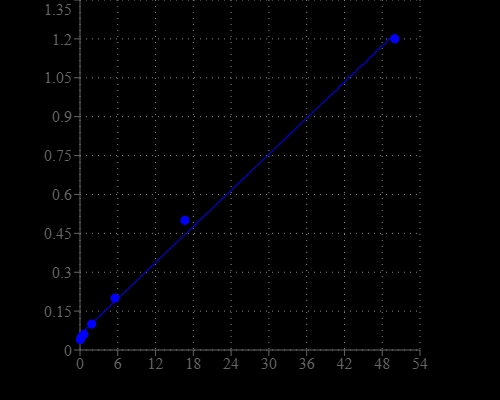
Phosphate dose response was measured with PhosphoWorks™ Colorimetric MESG Phosphate Assay Kit on a 96-well UV plate using a SpectraMax Plus microplate reader (Molecular Devices).