Products
Services
Resources
Selection Guides
About
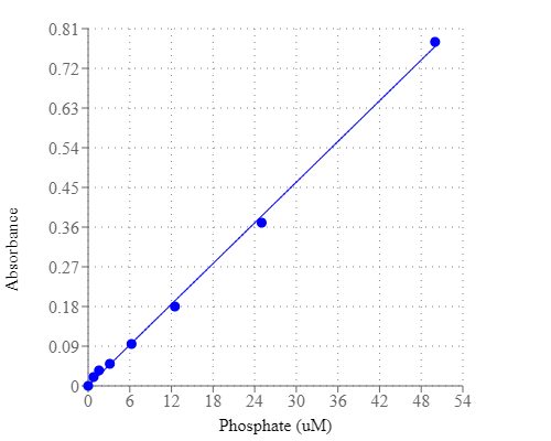
PhosphoWorks™ Colorimetric Phosphate Assay Kit
Blue Color
Phosphate is involved in many biological reactions. For example, phosphatases, ATPases and several other enzymes catalyze reactions in which inorganic phosphate (Pi) is released from a substrate. This PhosphoWorks™ Phosphate Assay Kit has been developed for measuring the activity of any Pi-generating enzyme. The kit is formulated to give sensitive detection of Pi, providing an alternative to hazardous radioactive methods and other less sensitive colorimetric assays. The measurement of Pi is based on the change in absorbance of a malachite green derivative in the presence of molybdate. Unlike other malachite dye formulations, this kit gives a completely stable end-point signal that is not prone to precipitation.
| Catalog | Size | Price | Quantity |
|---|---|---|---|
| 21665 | 1000 Tests | Price |
Usage and storage
| Intended use | Research Use Only (RUO) |
Instrument settings
| Spectrophotometer | |
| Absorbance | 600 - 660 nm |
| Recommended plate | Clear bottom |
| Absorbance microplate reader | |
| Absorbance | 600 - 660 nm |
| Recommended plate | Clear bottom |
Contact us
| Telephone | |
| Fax | |
| sales@aatbio.com | |
| International | See distributors |
| Bulk request | Inquire |
| Custom size | Inquire |
| Technical Support | Contact us |
| Request quotation | Request |
| Purchase order | Send to sales@aatbio.com |
| Shipping | Standard overnight for United States, inquire for international |
Page updated on July 16, 2026