Portelite™ Fluorimetric ssDNA Quantitation Kit *Optimized for Cytocite™ and Qubit™ Fluorometers*
Ordering information
Price | |
Catalog Number | |
Unit Size | |
Quantity |
Additional ordering information
Telephone | 1-800-990-8053 |
Fax | 1-800-609-2943 |
sales@aatbio.com | |
International | See distributors |
Bulk request | Inquire |
Custom size | Inquire |
Shipping | Standard overnight for United States, inquire for international |
Spectral properties
Excitation (nm) | 498 |
Emission (nm) | 519 |
Storage, safety and handling
H-phrase | H303, H313, H333 |
Hazard symbol | XN |
Intended use | Research Use Only (RUO) |
R-phrase | R20, R21, R22 |
UNSPSC | 12171501 |
Related products
Overview | SDSProtocol |
Excitation (nm) 498 | Emission (nm) 519 |
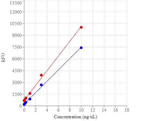
Portelite™ Fluorimetric ssDNA Quantitation Kit is designed to rapidly measure single-stranded DNA. The kit has all the essential reagents, including Helixyte™ Green ssDNA reagent, dilution buffer, and prediluted DNA standards. Helixyte™ Green ssDNA reagent is a sensitive fluorescent nucleic acid probe for quantifying oligonucleotides and single-stranded DNA (ssDNA) in solution. Simply dilute the reagent using the buffer provided, add your sample (any volume from 1–20 μL is acceptable), then read the concentration using CytoCite, Qubit® or other hand-held or desk top fluorometers (Qubit® is the trademark of ThermoFisher). The assay is accurate for initial sample concentrations from 50 pg/µL to 200 ng/µL, providing an assay range of 1–200 ng. The assay detects long oligonucleotides or ssDNA. Nucleotides and short oligo nucleotides of six bases or less do not interfere in the quantitation assay. The detection limit is not significantly interfered by the common contaminants in nucleic acid preparations, including salts, urea, ethanol, chloroform, detergents, proteins, nucleotides and short oligonucleotides of six bases. However, double-stranded DNA (dsDNA) and RNA do interfere with the assay as Helixyte™ Green ssDNA reagent binds to dsDNA and RNA to generate additional fluorescence signal. Portelite™ Fluorimetric ssDNA Quantitation Kit is optimized for CytoCite™ and Qubit® fluorometers.
Platform
Qubit Fluorometer
Excitation | 480 nm |
Emission | 530 nm |
Instrument specification(s) | 0.2 mL PCR vial |
CytoCite Fluorometer
Excitation | 480 nm |
Emission | 530 nm |
Instrument specification(s) | 0.2 mL PCR vial |
Components
Example protocol
AT A GLANCE
Protocol summary
- Prepare the Helixyte™ Green ssDNA working solution
- Add 190 µL of 1X Helixyte™ Green ssDNA working solution into each 0.2 mL PCR tube
- Add 10 µL of ssDNA Standards or test samples into each tube
- Incubate at room temperature for 2 minutes
- Monitor the fluorescence intensity with CytoCite™ fluorometer or Qubit™ fluorometer
Important
All kit components must be brought to room temperature before starting the experiment.PREPARATION OF WORKING SOLUTION
Helixyte™ Green ssDNA working solution
Make a 200-fold dilution of Helixyte™ Green ssDNA reagent (Component A) with Assay Buffer (Component B). For example, to prepare enough working solution for 5 samples, add 5 μL of Helixyte™ Green ssDNA (Component A) into 1 mL of Assay Buffer (Component B).Note Protect the working solution from light by covering it with foil or placing it in the dark. It’s recommended to prepare the solution in a plastic container rather than a glass container, as the dye may adsorb to the glass surface. For best results, this solution should be used within a few hours after the dilution.
SAMPLE EXPERIMENTAL PROTOCOL
The acceptable range for the sample volume could be 1~20 μL depending on the estimated concentration of the Nucleic Acid sample.
The following protocol is generated based on a sample volume of 10 µL with the DNA concentration in the range of 20~1000 ng /mL.
- Add 190 µL of 1X Helixyte™ Green ssDNA working solution into each Cytocite™ sample tube (#CCT100) or the equivalent 0.2 mL PCR tube.
Note Use thin-wall, polypropylene, clear 0.2 mL PCR tubes such as AAT Cat#CCT100. - Add 10 µL of ssDNA Standards or test samples into each tube, and then mix by vortexing for 2~3 seconds.
- Incubate all tubes at room temperature for 2 minutes.
- Insert the samples into CytoCite™ or Qubit™ and monitor the fluorescence intensity with the green fluorescence channel. Follow the appropriate procedures for the CytoCite™ Fluorometer. See the link below for detailed instructions: https://devices.aatbio.com/documentation/user-manual-for-cytocite-fluorometer
Preparation Of Standard Calibration Curve
- Perform a 1:2 serial dilution: Add 10 ng/μL ssDNA Standard #2 (Component D) into Assay Buffer (Component B) to get 10, 5, 2.5, 1.25, 0.62, 0.31, 0.15 ng/μL DNA standard dilutions.
- Add 190 µL of the Helixyte™ Green ssDNA working solution into each tube.
- Add 10 µL of standards into a 0.2 mL PCR tube and then mix by vortexing for 2∼3 seconds.
- Incubate the reaction at room temperature for 2 minutes.
- Insert the samples into CytoCite™ and monitor the fluorescence intensity with the green fluorescence channel.
Product Family
Name | Excitation (nm) | Emission (nm) |
Portelite™ Fluorimetric RNA Quantitation Kit*Optimized for Cytocite™ and Qubit™ Fluorometers* | 509 | 527 |
Images
References
View all 24 references: Citation Explorer
An OliGreen-responsive fluorescence sensor for sensitive detection of organophosphorus pesticide based on its specific selectivity towards T-Hg2+-T DNA structure.
Authors: Zhou, Xiaoyuan and Wang, Chenchen and Wu, Lina and Wei, Wei and Liu, Songqin
Journal: Spectrochimica acta. Part A, Molecular and biomolecular spectroscopy (2021): 119155
Authors: Zhou, Xiaoyuan and Wang, Chenchen and Wu, Lina and Wei, Wei and Liu, Songqin
Journal: Spectrochimica acta. Part A, Molecular and biomolecular spectroscopy (2021): 119155
A novel detection of radon based on its decay product inducing conformational changes of an aptamer probe.
Authors: Long, Minzhi and Deng, Han and Tian, Gang and Song, Chunli and Liu, Hongwen and Shen, Yi and Lv, Changyin
Journal: Analytica chimica acta (2016): 202-7
Authors: Long, Minzhi and Deng, Han and Tian, Gang and Song, Chunli and Liu, Hongwen and Shen, Yi and Lv, Changyin
Journal: Analytica chimica acta (2016): 202-7
Studies of DNA Aptamer OliGreen and PicoGreen Fluorescence Interactions in Buffer and Serum.
Authors: Bruno, John G and Sivils, Jeffrey C
Journal: Journal of fluorescence (2016): 1479-87
Authors: Bruno, John G and Sivils, Jeffrey C
Journal: Journal of fluorescence (2016): 1479-87
Accumulation of single-stranded DNA in Escherichia coli carrying the colicin plasmid pColE3-CA38.
Authors: Morales, Magali and Attai, Hedieh and Troy, Kimberly and Bermudes, David
Journal: Plasmid (2015): 7-16
Authors: Morales, Magali and Attai, Hedieh and Troy, Kimberly and Bermudes, David
Journal: Plasmid (2015): 7-16
Probing the inherent stability of siRNA immobilized on nanoparticle constructs.
Authors: Barnaby, Stacey N and Lee, Andrew and Mirkin, Chad A
Journal: Proceedings of the National Academy of Sciences of the United States of America (2014): 9739-44
Authors: Barnaby, Stacey N and Lee, Andrew and Mirkin, Chad A
Journal: Proceedings of the National Academy of Sciences of the United States of America (2014): 9739-44
Cost-effective and scalable DNA extraction method from dried blood spots.
Authors: Saavedra-Matiz, Carlos A and Isabelle, Jason T and Biski, Chad K and Duva, Salvatore J and Sweeney, Melissa L and Parker, April L and Young, Allison J and Diantonio, Lisa L and Krein, Lea M and Nichols, Matthew J and Caggana, Michele
Journal: Clinical chemistry (2013): 1045-51
Authors: Saavedra-Matiz, Carlos A and Isabelle, Jason T and Biski, Chad K and Duva, Salvatore J and Sweeney, Melissa L and Parker, April L and Young, Allison J and Diantonio, Lisa L and Krein, Lea M and Nichols, Matthew J and Caggana, Michele
Journal: Clinical chemistry (2013): 1045-51
Sensitive fluorescence assay of anthrax protective antigen with two new DNA aptamers and their binding properties.
Authors: Oh, Byul Nim and Lee, Sungeun and Park, Hye-Yeon and Baeg, Jin-Ook and Yoon, Moon-Young and Kim, Jinheung
Journal: The Analyst (2011): 3384-8
Authors: Oh, Byul Nim and Lee, Sungeun and Park, Hye-Yeon and Baeg, Jin-Ook and Yoon, Moon-Young and Kim, Jinheung
Journal: The Analyst (2011): 3384-8
Enrichment and fluorescence enhancement of adenosine using aptamer-gold nanoparticles, PDGF aptamer, and Oligreen.
Authors: Chen, Shih-Ju and Huang, Chih-Ching and Chang, Huan-Tsung
Journal: Talanta (2010): 493-8
Authors: Chen, Shih-Ju and Huang, Chih-Ching and Chang, Huan-Tsung
Journal: Talanta (2010): 493-8
Fluorescently imaged particle counting immunoassay for sensitive detection of DNA modifications.
Authors: Wang, Zhixin and Wang, Xiaoli and Liu, Shengquan and Yin, Junfa and Wang, Hailin
Journal: Analytical chemistry (2010): 9901-8
Authors: Wang, Zhixin and Wang, Xiaoli and Liu, Shengquan and Yin, Junfa and Wang, Hailin
Journal: Analytical chemistry (2010): 9901-8
Analysis of DNA complexes with small solutes by CE with LIF detection.
Authors: Lee, Kun-Hong and Huang, Ming-Feng and Liu, Chi-Wei and Chang, Huan-Tsung
Journal: Electrophoresis (2010): 1101-7
Authors: Lee, Kun-Hong and Huang, Ming-Feng and Liu, Chi-Wei and Chang, Huan-Tsung
Journal: Electrophoresis (2010): 1101-7
Application notes
A Novel Fluorescent Probe for Imaging and Detecting Hydroxyl Radical in Living Cells
Fluorescent Oligonucleotide Labeling Reagents
Monitoring of Mitochondrial Membrane Potential Changes in Live Cells Using JC-10
Selective Analysis of RNA in Live and Fixed Cells with StrandBrite RNA Green
Cell Loading Protocol For Fluorescent pH Indicator, BCECF-AM
Fluorescent Oligonucleotide Labeling Reagents
Monitoring of Mitochondrial Membrane Potential Changes in Live Cells Using JC-10
Selective Analysis of RNA in Live and Fixed Cells with StrandBrite RNA Green
Cell Loading Protocol For Fluorescent pH Indicator, BCECF-AM
FAQ
I ordered your phalloidin-amine (Cat #5302) so I can conjugate it to my oligo. Do you have a recommended protocol I can use?
What dye works best for staining and tracking lysosomes in live cells for several hours?
How can I lyse my cells without lysing the nuclear membrane?
Do you have any dual-fluorescence nucleic acid stains that interact with both DNA and RNA?
Do you have any fixable mitochondria staining assay kits?
What dye works best for staining and tracking lysosomes in live cells for several hours?
How can I lyse my cells without lysing the nuclear membrane?
Do you have any dual-fluorescence nucleic acid stains that interact with both DNA and RNA?
Do you have any fixable mitochondria staining assay kits?