Products
Services
Resources
Selection Guides
About
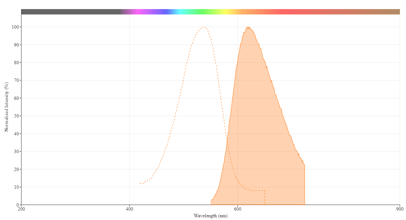
Propidium iodide
1 mg/mL aqueous solution
Propidium Iodide (1 mg/mL) is a DNA-intercalating red fluorescent dye that selectively stains dead cells with compromised membranes, widely used for viability assessment in flow cytometry and fluorescence microscopy.
- DNA-intercalating red fluorophore: Propidium iodide inserts between DNA base pairs and fluoresces intensely when bound (excitation ≈ 537 nm, emission ≈ 618 nm).
- Selective dead-cell marker: Its double positive charge keeps it out of intact membranes, so it stains only cells with compromised membranes, making it ideal for viability assays.
- Flow-cytometry friendly: Efficiently excited by the common 488 nm laser and pairs seamlessly with dyes like acridine orange or Annexin V-FITC for multi-parameter analyses.
| Catalog | Size | Price | Quantity |
|---|---|---|---|
| 17585 | 5 mL | Price | |
| 17586 | 10 mL | Price |

Physical properties
| Molecular weight | 668.39 |
| Solvent | Water |
Spectral properties
| Extinction coefficient (cm -1 M -1) | 6000 1 |
| Excitation (nm) | 537 |
| Emission (nm) | 618 |
| Quantum yield | 0.2 1 |
Storage, safety and handling
| H-phrase | H303, H313, H333 |
| Hazard symbol | XN |
| Intended use | Research Use Only (RUO) |
| R-phrase | R20, R21, R22 |
| Storage | Freeze (< -15 °C); Minimize light exposure |
| CAS | 25535-16-4 |
Contact us
| Telephone | |
| Fax | |
| sales@aatbio.com | |
| International | See distributors |
| Bulk request | Inquire |
| Custom size | Inquire |
| Technical Support | Contact us |
| Request quotation | Request |
| Purchase order | Send to sales@aatbio.com |
| Shipping | Standard overnight for United States, inquire for international |
Page updated on September 16, 2024