Products
Services
Resources
Selection Guides
About
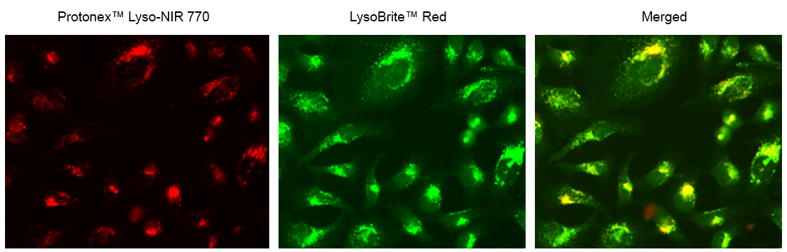
Protonex™ Lyso-NIR 770
Protonex™ Lyso-NIR 770 is a fluorescent derivative prepared from our Protonex™ NIR 770 NHS ester. It is cell-permeable and selectively localized in acidic organelles such as lysosomes. It is the only cell-permeable acidotropic pH dye with NIR fluorescence for monitoring pH changes in acidic organelles. Its fluorescence can be readily captured with the common Cy7 filter set that is equipped with most of the fluorescence instruments. Due to its significantly red-shifted excitation and emission wavelengths, it has much lower background than the existing pH sensors (such as Lysosensors). It might be used in vivo for monitoring pH changes in animals due to the significantly improved skin-penetration capability of NIR excitation.
| Catalog | Size | Price | Quantity |
|---|---|---|---|
| 21242 | 1 mg | Price |

Physical properties
| Molecular weight | 736.76 |
| Solvent | DMSO |
Spectral properties
| Excitation (nm) | 748 |
| Emission (nm) | 769 |
Storage, safety and handling
| H-phrase | H303, H313, H333 |
| Hazard symbol | XN |
| Intended use | Research Use Only (RUO) |
| R-phrase | R20, R21, R22 |
| Storage | Freeze (< -15 °C); Minimize light exposure |
Contact us
| Telephone | |
| Fax | |
| sales@aatbio.com | |
| International | See distributors |
| Bulk request | Inquire |
| Custom size | Inquire |
| Technical Support | Contact us |
| Request quotation | Request |
| Purchase order | Send to sales@aatbio.com |
| Shipping | Standard overnight for United States, inquire for international |
Page updated on July 14, 2026