Products
Services
Resources
Selection Guides
About
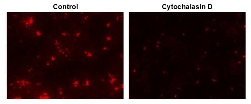
Protonex™ Red 600-E. coli Conjugate
The Protonex™ Red 600–E. coli Conjugate is a ready-to-use, pH-sensitive fluorescent tool for monitoring phagocytosis and intracellular acidification processes in live cells.
- pH-activated Fluorescence: Non-fluorescent at neutral pH and becomes strongly fluorescent in acidic compartments such as phagosomes and phagolysosomes
- E. coli-based Targeting: E. coli particles serve as biologically relevant substrates that mimic bacterial infection and are efficiently recognized by phagocytes
- Multipurpose Reagent: Suitable for incorporation into custom assay workflows for microscopy, flow cytometry, or plate-based readouts
| Catalog | Size | Price | Quantity |
|---|---|---|---|
| 21176 | 100 Tests | Price |

Physical properties
| Solvent |
Spectral properties
| Excitation (nm) | 576 |
| Emission (nm) | 597 |
Usage and storage
| Intended use | Research Use Only (RUO) |
| Storage | Refrigerated (2-8 °C); Minimize light exposure |
Instrument settings
| Fluorescence microscope | |
| Excitation | TRITC |
| Emission | TRITC |
| Recommended plate | Black wall/clear bottom |
| Fluorescence microplate reader | |
| Excitation | 540nm |
| Emission | 590nm |
| Recommended plate | Black wall/clear bottom |
| Instrument specification(s) | Bottom read mode |
Contact us
| Telephone | |
| Fax | |
| sales@aatbio.com | |
| International | See distributors |
| Bulk request | Inquire |
| Custom size | Inquire |
| Technical Support | Contact us |
| Request quotation | Request |
| Purchase order | Send to sales@aatbio.com |
| Shipping | Standard overnight for United States, inquire for international |
Page updated on July 15, 2026