logo
AAT Bioquest

ReadiLink™ Rapid Cy3 Antibody Labeling Kit *Microscale Optimized for Labeling 50 μg Antibody Per Reaction*

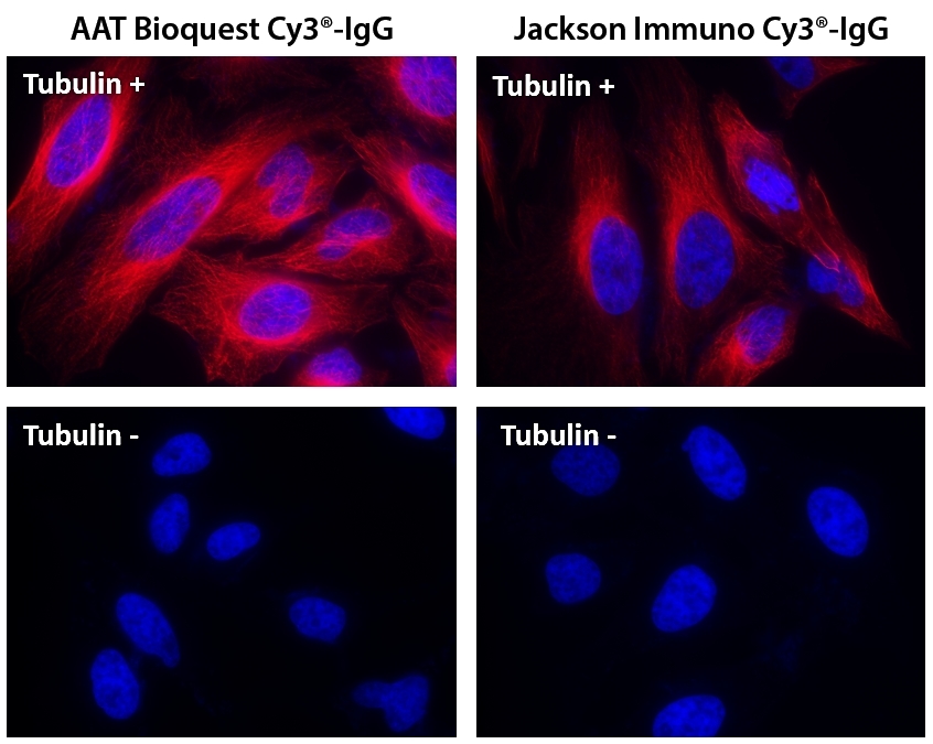
HeLa cells were incubated with (Tubulin+) or without (Tubulin-) mouse anti-tubulin followed by AAT&rsquo;s Cy3<sup>&reg;</sup>&nbsp;goat anti-mouse IgG conjugate (Red, Left) or Jackson&rsquo;s goat anti-mouse IgG conjugated with Cy3<sup>&reg;</sup>&nbsp; (Red, Right), respectively. Cell nuclei were stained with Hoechst 33342 (Blue, Cat# 17530).
Ordering information
Price
Catalog Number
Unit Size
Quantity
Add to cart
Additional ordering information
Telephone1-800-990-8053
Fax1-800-609-2943
Emailsales@aatbio.com
InternationalSee distributors
Bulk requestInquire
Custom sizeInquire
ShippingStandard overnight for United States, inquire for international
Request quotation
Spectral properties
Correction Factor (260 nm)0.07
Correction Factor (280 nm)0.073
Extinction coefficient (cm -1 M -1)1500001
Excitation (nm)555
Emission (nm)569
Quantum yield0.151
Storage, safety and handling
H-phraseH303, H313, H333
Hazard symbolXN
Intended useResearch Use Only (RUO)
R-phraseR20, R21, R22
UNSPSC12171501
Related products
ReadiLink™ Rapid mFluor™ Violet 450 Antibody Labeling Kit *Microscale Optimized for Labeling 50 μg Antibody Per Reaction*
ReadiLink™ Rapid mFluor™ Violet 420 Antibody Labeling Kit *Microscale Optimized for Labeling 50 μg Antibody Per Reaction*
ReadiLink™ Rapid mFluor™ Violet 510 Antibody Labeling Kit *Microscale Optimized for Labeling 50 μg Antibody Per Reaction*
ReadiLink™ Rapid mFluor™ Violet 540 Antibody Labeling Kit *Microscale Optimized for Labeling 50 μg Antibody Per Reaction*
ReadiLink™ Rapid mFluor™ Blue 570 Antibody Labeling Kit *Microscale Optimized for Labeling 50 μg Antibody Per Reaction*
ReadiLink™ Rapid mFluor™ Green 620 Antibody Labeling Kit *Microscale Optimized for Labeling 50 μg Antibody Per Reaction*
ReadiLink™ Rapid mFluor™ Yellow 630 Antibody Labeling Kit *Microscale Optimized for Labeling 50 μg Antibody Per Reaction*
ReadiLink™ Rapid mFluor™ Red 700 Antibody Labeling Kit *Microscale Optimized for Labeling 50 μg Antibody Per Reaction*
ReadiLink™ Rapid mFluor™ Red 780 Antibody Labeling Kit *Microscale Optimized for Labeling 50 μg Antibody Per Reaction*
ReadiLink™ Rapid iFluor® 350 Antibody Labeling Kit *Microscale Optimized for Labeling 50 μg Antibody Per Reaction*
ReadiLink™ Rapid iFluor® 594 Antibody Labeling Kit *Microscale Optimized for Labeling 50 μg Antibody Per Reaction*
ReadiLink™ Rapid iFluor® 647 Antibody Labeling Kit *Microscale Optimized for Labeling 50 μg Antibody Per Reaction*
ReadiLink™ Rapid iFluor® 680 Antibody Labeling Kit *Microscale Optimized for Labeling 50 μg Antibody Per Reaction*
ReadiLink™ Rapid iFluor® 700 Antibody Labeling Kit *Microscale Optimized for Labeling 50 μg Antibody Per Reaction*
ReadiLink™ Rapid iFluor® 750 Antibody Labeling Kit *Microscale Optimized for Labeling 50 μg Antibody Per Reaction*
ReadiLink™ Rapid iFluor® 488 Antibody Labeling Kit *Microscale Optimized for Labeling 50 μg Antibody Per Reaction*
ReadiLink™ Rapid iFluor® 633 Antibody Labeling Kit *Microscale Optimized for Labeling 50 μg Antibody Per Reaction*
ReadiLink™ Rapid iFluor® 790 Antibody Labeling Kit *Microscale Optimized for Labeling 50 μg Antibody Per Reaction*
ReadiLink™ Rapid trFluor™ Eu Antibody Labeling Kit *Microscale Optimized for Labeling 50 ug Antibody Per Reaction*
ReadiLink™ Rapid trFluor™ Tb Antibody Labeling Kit *Microscale Optimized for Labeling 50 ug Antibody Per Reaction*
ReadiLink™ Protein Conjugation Stop Buffer
ReadiLink™ BSA Conjugation Kit
ReadiLink™ KLH Conjugation Kit
ReadiLink™ Protein Biotinylation Kit
ReadiLink™ Protein Biotinylation Kit *Powered by ReadiView™ Biotin Visionization Technology*
ReadiLink™ xtra Rapid iFluor® 350 Antibody Labeling Kit *BSA-Compatible*
ReadiLink™ xtra Rapid iFluor® 488 Antibody Labeling Kit *BSA-Compatible*
ReadiLink™ xtra Rapid iFluor® 555 Antibody Labeling Kit *BSA-Compatible*
ReadiLink™ xtra Rapid iFluor® 594 Antibody Labeling Kit *BSA-Compatible*
ReadiLink™ xtra Rapid iFluor® 647 Antibody Labeling Kit *BSA-Compatible*
ReadiLink™ xtra Rapid iFluor® 750 Antibody Labeling Kit *BSA-Compatible*
ReadiLink™ xtra Rapid FITC Antibody Labeling Kit *BSA-Compatible*
ReadiLink™ xtra Rapid Cy3 Antibody Labeling Kit *BSA-Compatible*
ReadiLink™ xtra Rapid Cy5 Antibody Labeling Kit *BSA-Compatible*
ReadiLink™ xtra Rapid Cy7 Antibody Labeling Kit *BSA-Compatible*
ReadiLink™ xtra Rapid XFD594 Antibody Labeling Kit *BSA-Compatible, XFD594 Same Structure to Alexa Fluor™ 594*
ReadiLink™ iFluor® 488 Nick Translation dsDNA Labeling Kit
ReadiLink™ iFluor® 555 Nick Translation dsDNA Labeling Kit
ReadiLink™ iFluor® 647 Nick Translation dsDNA Labeling Kit
ReadiLink™ Cy3 Nick Translation dsDNA Labeling Kit
ReadiLink™ Cy5 Nick Translation dsDNA Labeling Kit
ReadiLink™ Biotin Nick Translation dsDNA Labeling Kit
ReadiLink™ DIG (Digoxigenin) Nick Translation dsDNA Labeling Kit
ReadiLink™ iFluor® 488 Oligo and ssDNA Labeling Kit
ReadiLink™ iFluor® 555 Oligo and ssDNA Labeling Kit
ReadiLink™ iFluor® 647 Oligo and ssDNA Labeling Kit
ReadiLink™ Cy3 Oligo and ssDNA Labeling Kit
ReadiLink™ Cy5 Oligo and ssDNA Labeling Kit
ReadiLink™ Biotin Oligo and ssDNA Labeling Kit
ReadiLink™ DIG (Digoxigenin) Oligo and ssDNA Labeling Kit
ReadiLink™ xtra Rapid XFD488 Antibody Labeling Kit *BSA-Compatible, XFD488 Same Structure to Alexa Fluor™ 488*
ReadiLink™ xtra Rapid XFD555 Antibody Labeling Kit *BSA-Compatible, XFD555 Same Structure to Alexa Fluor™ 555*
ReadiLink™ xtra Rapid XFD647 Antibody Labeling Kit *BSA-Compatible, XFD647 Same Structure to Alexa Fluor™ 647*
ReadiLink™ xtra Rapid XFD750 Antibody Labeling Kit *BSA-Compatible, XFD750 Same Structure to Alexa Fluor™ 750*
ReadiLink™ iFluor® 488 FISH Fluorescence Imaging Kit
ReadiLink™ iFluor® 555 FISH Fluorescence Imaging Kit
ReadiLink™ iFluor® 647 FISH Fluorescence Imaging Kit
ReadiLink™ Rapid iFluor® 350 Antibody Labeling Kit *Production Scale*
ReadiLink™ Rapid iFluor® 488 Antibody Labeling Kit *Production Scale*
ReadiLink™ Rapid iFluor® 594 Antibody Labeling Kit *Production Scale*
ReadiLink™ Rapid iFluor® 647 Antibody Labeling Kit *Production Scale*
ReadiLink™ Rapid iFluor® 750 Antibody Labeling Kit *Production Scale*
ReadiLink™ Psoralen-A17-Biotin
ReadiLink™ Rapid mFluor™ Violet 610 Antibody Labeling Kit *Microscale Optimized for Labeling 50 µg Antibody Per Reaction*
Show More (54)

OverviewpdfSDSpdfProtocol


Correction Factor (260 nm)
0.07
Correction Factor (280 nm)
0.073
Extinction coefficient (cm -1 M -1)
1500001
Excitation (nm)
555
Emission (nm)
569
Quantum yield
0.151
Cy3 is one of the most popular fluorescent labeling dyes for preparing orange-red fluorescent bioconjugates. However, most of the commercial Cy3 labeling kits require intensive hands-on time. This Cy3 ReadiLink™ labeling kit is one of the most robust protein labeling kits for preparing Cy3-labeled antibody conjugates or other protein conjugates. It essentially only requires 2 simple mixing steps without a column purification required. The kit provides all the essential components for labeling ~2x50 ug antibody. Each of the two vials of Cy3 dye provided in the kit is optimized for labeling ~50 µg antibody. This Cy3 protein labeling kit provides a convenient method to label monoclonal, polyclonal antibodies or other proteins (>10 kDa).


readilinkworkflow


Figure 1. Overview of the ReadiLink™ Rapid Antibody Labeling protocol. In just two simple steps, and with no purification necessary, covalently label microgram amounts of antibodies in under an hour.
Components


Example protocol


AT A GLANCE

Important
Warm all the components and centrifuge the vials briefly before opening, and immediately prepare the required solutions before starting your conjugation. The following protocol is for recommendation.

PREPARATION OF WORKING SOLUTION

Protein working solution (Solution A)
For labeling 50 µg of protein (assuming the target protein concentration is 1 mg/mL), mix 5 µL (10% of the total reaction volume) of Reaction Buffer (Component B) with 50 µL of the target protein solution.
Note     If you have a different protein concentration, adjust the protein volume accordingly to make ~50 µg of protein available for your labeling reaction.
Note     For labeling 100 µg of protein (assuming the target protein concentration is 1 mg/mL), mix 10 µL (10% of the total reaction volume) of Reaction Buffer (Component B) with 100 µL of the target protein solution.
Note     The protein should be dissolved in 1X phosphate buffered saline (PBS), pH 7.2 - 7.4; if the protein is dissolved in glycine buffer, it must be dialyzed against 1X PBS, pH 7.2 - 7.4, or use Amicon Ultra-0.5, Ultracel-10 Membrane, 10 kDa (cat# UFC501008 from Millipore) to remove free amines or ammonium salts (such as ammonium sulfate and ammonium acetate) that are widely used for protein precipitation.
Note     Impure antibodies or antibodies stabilized with bovine serum albumin (BSA) or gelatin will not be labeled well.
Note     For optimal labeling efficiency, a final protein concentration range of 1 - 2 mg/mL is recommended, with a significantly reduced conjugation efficiency at less than 1 mg/mL.

SAMPLE EXPERIMENTAL PROTOCOL

Run conjugation reaction
  1. Add the protein working solution (Solution A) to ONE vial of labeling dye (Component A), and mix them well by repeatedly pipetting for a few times or vortex the vial for a few seconds.
    Note     If labeling 100 µg of protein, use both vials (Component A) of labeling dye by dividing the 100 µg of protein into 2 x 50 µg of protein and reacting each 50 µg of protein with one vial of labeling dye. Then combine both vials for the next step.
  2. Keep the conjugation reaction mixture at room temperature for 30 - 60 minutes.
    Note     The conjugation reaction mixture can be rotated or shaken for longer time if desired. 

Stop Conjugation reaction
  1. Add 5 µL (for 50 µg protein) or 10 µL (for 100 µg protein) which is 10% of the total reaction volume of TQ™-Dyed Quench Buffer (Component C) into the conjugation reaction mixture; mix well.
  2. Incubate at room temperature for 10 minutes. The labeled protein (antibody) is now ready to use. 

Storage of Protein Conjugate
The protein conjugate should be stored at > 0.5 mg/mL in the presence of a carrier protein (e.g., 0.1% bovine serum albumin). For longer storage, the protein conjugates could be lyophilized or divided into single-used aliquots and stored at ≤ –20°C.

Spectrum


Open in Advanced Spectrum Viewer
spectrum

Spectral properties

Correction Factor (260 nm)0.07
Correction Factor (280 nm)0.073
Extinction coefficient (cm -1 M -1)1500001
Excitation (nm)555
Emission (nm)569
Quantum yield0.151

Product Family


NameExcitation (nm)Emission (nm)Extinction coefficient (cm -1 M -1)Quantum yieldCorrection Factor (280 nm)Correction Factor (260 nm)
ReadiLink™ Rapid FITC Antibody Labeling Kit *Microscale Optimized for Labeling 50 μg Antibody Per Reaction*491516730000.920.35-
ReadiLink™ Rapid Cy5 Antibody Labeling Kit *Microscale Optimized for Labeling 50 μg Antibody Per Reaction*65167025000010.271, 0.420.030.02
ReadiLink™ Rapid Cy7 Antibody Labeling Kit *Microscale Optimized for Labeling 50 μg Antibody Per Reaction*7567792500000.30.0360.05
ReadiLink™ Rapid XFD488 Antibody Labeling Kit *XFD488 Same Structure to Alexa Fluor™ 488*499520710000.9210.110.30
ReadiLink™ Rapid XFD555 Antibody Labeling Kit *XFD555 Same Structure to Alexa Fluor™ 555*5535681500000.110.080.08
ReadiLink™ Rapid XFD594 Antibody Labeling Kit *XFD594 Same Structure to Alexa Fluor™ 594*590618900000.6610.560.43
ReadiLink™ Rapid XFD647 Antibody Labeling Kit *XFD647 Same Structure to Alexa Fluor™ 647*6506712390000.3310.030.00
ReadiLink™ Rapid XFD750 Antibody Labeling Kit *XFD750 Same Structure to Alexa Fluor™ 750*7527762400000.1210.040.00
ReadiLink™ Rapid FITC Antibody Labeling Kit *Production Scale*491516730000.920.35-
ReadiLink™ Rapid Cy5 Antibody Labeling Kit *Production Scale*65167025000010.271, 0.420.030.02
ReadiLink™ Rapid XFD488 Antibody Labeling Kit *Production Scale*499520710000.9210.110.30
ReadiLink™ Rapid XFD555 Antibody Labeling Kit *Production Scale*5535681500000.110.080.08
ReadiLink™ Rapid XFD594 Antibody Labeling Kit *Production Scale*590618900000.6610.560.43
ReadiLink™ Rapid Cy7 Antibody Labeling Kit *Production Scale*7567792500000.30.0360.05
ReadiLink™ Rapid XFD647 Antibody Labeling Kit *Production Scale*6506712390000.3310.030.00
ReadiLink™ Rapid XFD750 Antibody Labeling Kit *Production Scale*7527762400000.1210.040.00
Show More (7)

Images


Citations


View all 14 citations: Citation Explorer
Affinity Peptide-Tethered Suspension Hydrogel Sensor for Selective and Sensitive Detection of Influenza Virus
Authors: Kim, Ji Hong and Jeong, Hye-Seon and Hwang, Jaehyeon and Kweon, Dae-Hyuk and Choi, Chang-Hyung and Park, Jong Pil
Journal: ACS Applied Materials \& Interfaces (2023)
Antibody-antibiotic conjugate targeted therapy for orthopedic implant-associated intracellular S. aureus infections
Authors: Hu, Ning and Zhang, Yanhao and Yang, Jianye and Zhao, Liqun and Zhang, Xiaokai and Yang, Yun and Zhang, Jinyong and Zou, Yinshuang and Wei, Keyu and Zhao, Chen and others,
Journal: (2023)
Thermo-sensitive hydrogel PLGA-PEG-PLGA as a vaccine delivery system for intramuscular immunization
Authors: Wang, Xiaoyan and Zhang, Yu and Xue, Wei and Wang, Hong and Qiu, Xiaozhong and Liu, Zonghua
Journal: Journal of Biomaterials Applications (2017): 923--932
Cube-shaped theranostic paclitaxel prodrug nanocrystals with surface functionalization of SPC and MPEG-DSPE for imaging and chemotherapy
Authors: Guo, Fuqiang and Shang, Jiajia and Zhao, Hai and Lai, Kangrong and Li, Yang and Fan, Zhongxiong and Hou, Zhenqing and Su, Guanghao
Journal: Colloids and Surfaces B: Biointerfaces (2017)
Light/magnetic hyperthermia triggered drug released from multi-functional thermo-sensitive magnetoliposomes for precise cancer synergetic theranostics
Authors: Guo, Yuxin and Zhang, Yang and Ma, Jinyuan and Li, Qi and Li, Yang and Zhou, Xinyi and Zhao, Dan and Song, Hua and Chen, Qing and Zhu, Xuan
Journal: Journal of Controlled Release (2017)
Molecular Basis and Consequences of the Cytochrome c-tRNA Interaction
Authors: Liu, Cuiping and Stonestrom, Aaron J and Christian, Thomas and Yong, Jeongsik and Takase, Ryuichi and Hou, Ya-Ming and Yang, Xiaolu
Journal: Journal of Biological Chemistry (2016): 10426--10436
Click-electron microscopy for imaging metabolically tagged nonprotein biomolecules
Authors: Ngo, John T and Adams, Stephen R and Deerinck, Thomas J and Boassa, Daniela and Rodriguez-Rivera, Frances and Palida, Sakina F and Bertozzi, Carolyn R and Ellisman, Mark H and Tsien, Roger Y
Journal: Nat Chem Biol (2016): 459--465
Design, synthesis and evaluation of VEGF-siRNA/CRS as a novel vector for gene delivery
Authors: Zhao, Wen and Zhang, Yifan and Jiang, Xueyun and Cui, Chunying
Journal: Drug Design, Development and Therapy (2016): 3851
Affinity-Controlled Protein Encapsulation into Sub-30 nm Telodendrimer Nanocarriers by Multivalent and Synergistic Interactions
Authors: Wang, Xu and Shi, Changying and Zhang, Li and Bodman, Alexa and Guo, D and an , undefined and Wang, Lili and Hall, Walter A and Wilkens, Stephan and Luo, Juntao
Journal: Biomaterials (2016)
Carboxymethyl Dextran-Stabilized Polyethylenimine-Poly (epsilon-caprolactone) Nanoparticles-Mediated Modulation of MicroRNA-34a Expression via Small-Molecule Modulator for Hepatocellular Carcinoma Therapy
Authors: Deng, Xiongwei and Yin, Zhaoxia and Zhou, Zhixiang and Wang, Yihui and Zhang, Fang and Hu, Qin and Yang, Yishu and Lu, Jianqing and Wu, Yan and Sheng, Wang and others, undefined
Journal: ACS applied materials &amp; interfaces (2016): 17068--17079

References


View all 21 references: Citation Explorer
Excitation of Cy5 in self-assembled lipid bilayers using optical microresonators
Authors: Freeman LM, Li S, Dayani Y, Choi HS, Malmstadt N, Armani AM.
Journal: Appl Phys Lett (2011): 143703
Theranostic cRGD-BioShuttle Constructs Containing Temozolomide- and Cy7 For NIR-Imaging and Therapy
Authors: Wiessler M, Hennrich U, Pipkorn R, Waldeck W, Cao L, Peter J, Ehemann V, Semmler W, Lammers T, Braun K.
Journal: Theranostics (2011): 381
Rational approach to select small peptide molecular probes labeled with fluorescent cyanine dyes for in vivo optical imaging
Authors: Berezin MY, Guo K, Akers W, Livingston J, Solomon M, Lee H, Liang K, Agee A, Achilefu S.
Journal: Biochemistry (2011): 2691
In vivo detection of embryonic stem cell-derived cardiovascular progenitor cells using Cy3-labeled Gadofluorine M in murine myocardium
Authors: Adler ED, Bystrup A, Briley-Saebo KC, Mani V, Young W, Giovanonne S, Altman P, Kattman SJ, Frank JA, Weinmann HJ, Keller GM, Fayad ZA.
Journal: JACC Cardiovasc Imaging (2009): 1114
Quantitative proteomics by fluorescent labeling of cysteine residues using a set of two cyanine-based or three rhodamine-based dyes
Authors: Volke D, Hoffmann R.
Journal: Electrophoresis (2008): 4516
Surface plasmon resonance-enhanced fluorescence implementation of a single-step competition assay: demonstration of fatty acid measurement using an anti-fatty acid monoclonal antibody and a Cy5-labeled fatty acid
Authors: Vareiro MM, Tranchant I, Maplin S, Zak K, Gani MM, Slevin CJ, Hailes HC, Tabor AB, Cameron PJ, Jenkins AT, Williams DE.
Journal: Anal Biochem (2008): 243
Thiazole orange and Cy3: improvement of fluorescent DNA probes with use of short range electron transfer
Authors: Menacher F, Rubner M, Berndl S, Wagenknecht HA.
Journal: J Org Chem (2008): 4263
Near-infrared fluorescence imaging of tumor integrin alpha v beta 3 expression with Cy7-labeled RGD multimers
Authors: Wu Y, Cai W, Chen X.
Journal: Mol Imaging Biol (2006): 226
Cy7-Bis-dipicolylamine-zinc
Authors: Leung K., undefined
Journal: In: Molecular Imaging and Contrast Agent Database (MICAD), Bethesda (MD). (2004)
Cy7-Tetrameric arginine-glycine-aspartic acid peptide
Authors: Cheng KT., undefined
Journal: In: Molecular Imaging and Contrast Agent Database (MICAD), Bethesda (MD). (2004)