logo
AAT Bioquest

ReadiPrep™ Lipopolysaccharide (LPS) Isolation Kit

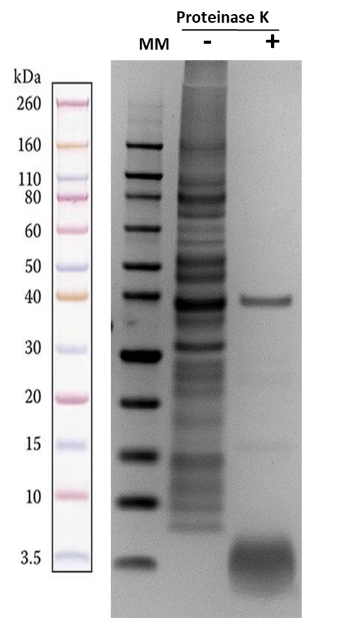
The ReadiPrep™ Lipopolysaccharide (LPS) Isolation Kit provides a safe and effective way to isolate LPS from the outer membrane of Gram-negative bacteria. It utilizes a bacterial membrane lysis buffer and enzymatic protein digestion to extract pure LPS from bacterial cultures, which can be quantified using carbohydrate detection methods. Unlike conventional approaches that rely on hazardous organic chemicals like chloroform or phenol, this kit provides a safer and more time-efficient alternative for isolating high-purity LPS for research applications. Lipopolysaccharides, or LPS, are found in the outer membrane of Gram-negative bacteria. It is a carbohydrate with a low molecular weight of 10-20 kDa. It is a complex molecule made of heterogeneous components comprising O antigen, core oligosaccharide, Lipid A, and non-carbohydrate components such as phosphate and amino acids groups. Lipid A, characterized by its multiple fatty acid chains, anchors the LPS into the bacterial membrane and contributes to the toxicity of the Gram-negative bacteria. Known as endotoxins, LPS is notorious for inducing potent inflammatory responses and sepsis when consumed by animals.

Example protocol

AT A GLANCE

Protocol Summary
  1. Grow bacteria on the LB agar plate.

  2. Add LPS isolation buffer to the bacterial pellet. Sonicate the mixture, then incubate it on ice for 10 minutes.
  3. Add proteinase K to the bacterial lysate and incubate the mixture at 60°C for one hour.

Important Note

Thaw all the kit components at room temperature before starting the experiment. 

SAMPLE EXPERIMENTAL PROTOCOL

  1. Grow the bacteria on an LB agar plate overnight at 37°C.

  2. The next day, collect bacterial colonies in ice-cold PBS. Measure the optical density (OD) at 600 nm using a spectrophotometer. Ensure the OD at 600 nm is greater than 0.6.

  3. Centrifuge the tube at 2500g for 10 minutes to collect the pellet. After removing the supernatant, centrifuge again at 2500g for 10 minutes to ensure all the supernatant is removed.

  4. Measured the weight of the pellets.

    Note: For an E. coli suspension with OD > 0.6, the pellet weight should be greater than 10 mg.

  5. Add 200 µL of isolation buffer to the LPS pellet. Use 200 µL for a 20 mg pellet.

  6. Sonicate the pellet three times for 30 seconds each at 10 watts. Then, incubate the tube on ice for 10 minutes.

  7. Centrifuge the tube at 2500g for 10 minutes at 4°C.

  8. Transfer the lysate into a new, clean tube.

  9. Add Proteinase K to the lysate at a concentration of 0.1 mg/mL.

    Note: For example, if you have 200 µL of lysate, add 1 µL of 20 mg/mL Proteinase K solution.

  10. Incubate the tube at 60 °C for one hour.

  11. Centrifuge the tube at 2500g for 10 minutes at 4°C.

  12. Collect the supernatant.

  13. LPS can be measured using carbohydrate detection methods. Alternatively, its purity can be assessed by running it on an SDS-PAGE gel and then staining it with Coomassie blue.

References

View all 50 references: Citation Explorer
Isolation and microbial transformation of tea sapogenin from seed pomace of Camellia oleifera with anti-inflammatory effects.
Authors: Shen, Pingping and Jiang, Xuewa and Zhang, Jingling and Wang, Jiayi and Raj, Richa and Li, Guolong and Ge, Haixia and Wang, Weiwei and Yu, Boyang and Zhang, Jian
Journal: Chinese journal of natural medicines (2024): 280-288
Influence of the isolation method on characteristics and functional activity of mesenchymal stromal cell-derived extracellular vesicles.
Authors: Malvicini, Ricardo and De Lazzari, Giada and Tolomeo, Anna Maria and Santa-Cruz, Diego and Ullah, Mujib and Cirillo, Carmine and Grumati, Paolo and Pacienza, Natalia and Muraca, Maurizio and Yannarelli, Gustavo
Journal: Cytotherapy (2024): 157-170
Isolation of New Diterpenoids from the Halophyte, Vitex rotundifolia, and their Antioxidant and Anti-inflammatory Activities.
Authors: Hwan Oh, Jung and Kong, Chang-Suk and Lee, Jihee and Kim, Eun-Hee and Seo, Youngwan
Journal: Chemistry & biodiversity (2024): e202301115
Isolation of Calenduloside E from Achyranthes bidentata Blume and its effects on LPS/D-GalN-induced acute liver injury in mice by regulating the AMPK-SIRT3 signaling pathway.
Authors: Guo, Pengli and Zeng, Mengnan and Liu, Meng and Zhang, Yuhan and Jia, Jufang and Zhang, Ziyu and Liang, Shulei and Zheng, Xiaoke and Feng, Weisheng
Journal: Phytomedicine : international journal of phytotherapy and phytopharmacology (2024): 155353
An Optimized Langendorff-free Method for Isolation and Characterization of Primary Adult Cardiomyocytes.
Authors: Nikouee, Azadeh and Yap, John Q and Rademacher, David J and Kim, Matthew and Zang, Qun Sophia
Journal: Research square (2024)
Page updated on June 10, 2025

Ordering information

Price
Unit size
Catalog Number60201
Quantity
Add to cart

Additional ordering information

Telephone1-800-990-8053
Fax1-800-609-2943
Emailsales@aatbio.com
InternationalSee distributors
Bulk requestInquire
Custom sizeInquire
Technical SupportContact us
Purchase orderSend to sales@aatbio.com
ShippingStandard overnight for United States, inquire for international
Request quotation

Storage, safety and handling

H-phraseH303, H313, H333
Hazard symbolXN
Intended useResearch Use Only (RUO)
R-phraseR20, R21, R22
UNSPSC12352200

Components

Bacterial lysate, prepared using the ReadiPrep™ Lipopolysaccharide (LPS) Isolation Kit, was boiled at 85 °C for 3-5 minutes in 1X loading buffer. The sample was then loaded onto a 4-12% SDS-PAGE gel and run for 70 minutes at 110V. Afterward, the gel was stained with Coomassie Blue to visualize the proteins.