logo
AAT Bioquest

ReadiPrep™ Mitochondrial/Cytoplasmic Fractionation Kit

Mitochondria fractions from HeLa cells were collected using ReadiPrep™ Mitochondrial/Cytoplasmic Fractionation Kit. The protein was quantified by Amplite® Fluorimetric Fluorescamine Protein Quantitation Kit (#11100). 100 µg of mitochondria fragment was incubated with or without MitoLite™ Green (#22675) and assayed with a NovoCyte 3000 flow cytometer.
Ordering information
Price
Catalog Number
Unit Size
Quantity
Add to cart
Additional ordering information
Telephone1-800-990-8053
Fax1-800-609-2943
Emailsales@aatbio.com
InternationalSee distributors
Bulk requestInquire
Custom sizeInquire
ShippingStandard overnight for United States, inquire for international
Request quotation
Storage, safety and handling
H-phraseH303, H313, H333
Hazard symbolXN
Intended useResearch Use Only (RUO)
R-phraseR20, R21, R22
UNSPSC12352200

OverviewpdfSDSpdfProtocol


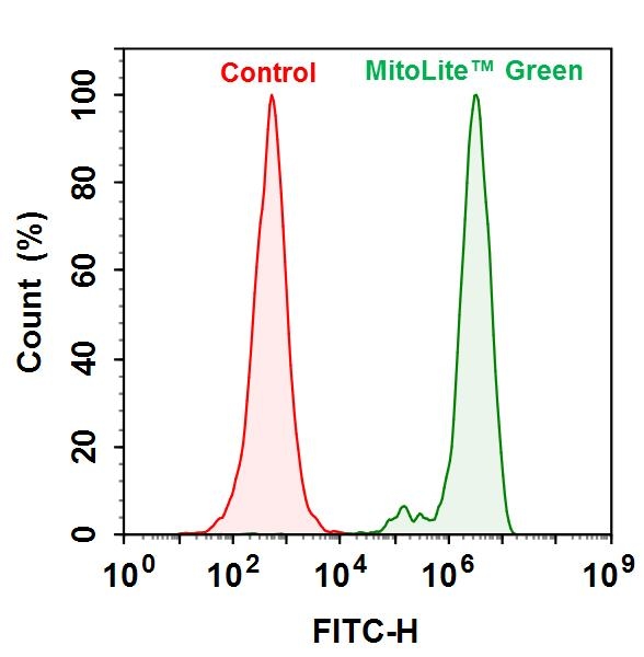
The ReadiPrep™ Mitochondrial/Cytoplasmic Fractionation Kit offers a convenient method for isolating mitochondria and cytoplasmic fractions from mammalian cells or tissue. The isolated mitochondria are intact and can be positively stained with a mitochondria marker (such as MitoLite™ Green). The isolated mitochondria preserve their biological activities and are compatible with many downstream applications including the study of mitochondrial respiration, mitochondria membrane potential, apoptosis, mtDAN and mtRNA, and mitochondrial protein profiling etc. The kit offers two options for the isolation of mitochondria. One option utilizes a reagent-based method which allows multiple sample preparation at the same time. The second option utilizes Dounce homogenization.

Components


Example protocol


AT A GLANCE

Protocol Summary
  1. Wash cells with PBS
  2. Add 2 mL of Isolation Buffer
  3. Incubate on ice for 10 minutes
  4. Homogenize the cells using Homogenizer or add Reagent A.
  5. Centrifuge at 7000 g for 10 mins
  6. Remove the supernatent (Cytoplasmic Fraction)
  7. Resuspend the pellets in Isolation Buffer
  8. Centrifuge at 7000 g for 10 minutes
  9. Resuspend the pellets in Assay Buffer (Mitochondria Fraction)
Important Note

Thaw all the kit components to room temperature before starting the experiment. Keep the buffer on ice during the experiment.

SAMPLE EXPERIMENTAL PROTOCOL

  1. Wash the cells once with cold PBS. For cells in suspension, collect the cells by centrifugation.
  2. Add 2 mL of Isolation Buffer (Component A) to cells and incubate on ice for 10 minutes.
  3. Two ways cytoplasmic fraction can be extraced. Option A: Add 40 µL of Reagent A (Component B) into the mix, vortex and incubate on ice for 10 minutes. Option B: Homogenize the cells using a Dounce Tissue Grinder by stroking 4-5 times.
  4. Centrifuge the sample at 600 g for 10 minutes at 4 °C and collect the supernatent and centrifuge again at 7,000 g for 10 minutes at 4 °C. The supernatent is cytoplasmic fraction.
  5. Re-suspend the pellet in 1 mL of Isolation Buffer (Component A) and centrifuge at 7,000 g for 10 minutes at 4 °C.
  6. Discard the supernatent, and resuspend the pellet in the appropriate assay buffer based on downstream applications.
  7. The protein concentration can be quantified by Amplite™ Fluorimetric Fluorescamine Protein Quantitation Kit (Cat# 11100).

Images


Citations


View all 2 citations: Citation Explorer
Involvement of Ferroptosis Induction and Oxidative Phosphorylation Inhibition in the Anticancer-Drug-Induced Myocardial Injury: Ameliorative Role of Pterostilbene
Authors: Fujii, Kiyomu and Fujiwara-Tani, Rina and Nukaga, Shota and Ohmori, Hitoshi and Luo, Yi and Nishida, Ryoichi and Sasaki, Takamitsu and Miyagawa, Yoshihiro and Nakashima, Chie and Kawahara, Isao and others,
Journal: International Journal of Molecular Sciences (2024): 3015
Phosphorylation of Cofilin-1 Enhances Paclitaxel Resistance of Epithelial Ovarian Cancer Cells by Inhibiting Apoptosis
Authors: Min, LI and Dong, Xu Dong and Lyu, Qiu Bo and Zhang, Wei and Huang, Shuai and Yang, Chun Xue and Di, CUI and Lai, Hui Ying
Journal: Biomedical and Environmental Sciences (2021): 465--477

References


View all 2 references: Citation Explorer
Studied by Rapid Fractionation of Barley (Hordeum vulgare) Protoplasts
Authors: Gardestrom, P.; Wigge, B., Influence of Photorespiration on ATP/ADP Ratios in the Chloroplasts, Mitochondria, and Cytosol, undefined
Journal: Plant Physiol (1988): 69-76
and mitochondria
Authors: Lilley, R. M.; Stitt, M.; Mader, G.; Heldt, H. W., Rapid fractionation of wheat leaf protoplasts using membrane filtration : the determination of metabolite levels in the chloroplasts, cytosol
Journal: Plant Physiol (1982): 965-70