logo
Products
Technologies
Applications
Services
Resources
Selection Guides
About
ReadiPrep™ Nuclear/Cytoplasmic Fractionation Kit
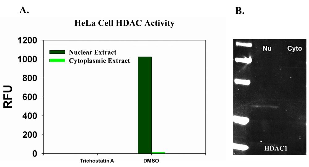
The ReadiPrep™ Nuclear/Cytoplasmic Fractionation Kit offers stepwise isolation of nuclear and cytoplasmic extract from mammalian cells or tissue. The isolated proteins have high concentrations and preserve their biological activities. The nuclear and cytoplasmic extracts from ReadiPrep™ Nuclear/Cytoplasmic Fractionation Kit are compatible with many downstream applications including enzyme activity assays and fluorescent Western blotting etc.
Nuclear and cytoplasmic extract from Hela cells were collected using ReadiPrep&trade; Nucleus/Cytosol Fractionation Kit (AAT Bioquest&reg;, Cat#60000). The protein was quantified by Amplite® Fluorimetric Fluorescamine Protein Quantitation Kit (AAT Bioquest&reg;, Cat#. 11100). <strong>A</strong>. 8 &micro;g of nuclear or cytoplasmic extract was incubated with or without HDAC inhibitor Trichostatin A, and HDAC activity was measured by Amplite® Fluorimetric HDAC Activity Assay Kit (AAT Bioquest&reg;, Cat#. 13601). <strong>B</strong>. 40 ug total protein of nuclear or cytoplasmic extract was used.&nbsp; 2 ug/ml of rabbit anti-HDAC1 antibody was used to probe the nitrocellulose membrane for overnight. 10 ug/ml of iFluorTM 647 goat anti-rabbit IgG (H+L) (AAT Bioquest&reg;, Cat#. 16809) was used. The detection was visualized by UVP MultiSpectral Imaging System (Biolite). M: Marker, Nu: Nuclear extract, Cyto: Cytoplasmic extract.
CatalogSize
Price
Quantity
6000050 Tests
Price
 
Storage, safety and handling

H-phraseH303, H313, H333
Hazard symbolXN
Intended useResearch Use Only (RUO)
R-phraseR20, R21, R22
UNSPSC12352200
Contact us

Telephone
Fax
Emailsales@aatbio.com
InternationalSee distributors
Bulk requestInquire
Custom sizeInquire
Technical SupportContact us
Request quotationRequest
Purchase orderSend to sales@aatbio.com
ShippingStandard overnight for United States, inquire for international
Page updated on September 25, 2025