Products
Services
Resources
Selection Guides
About
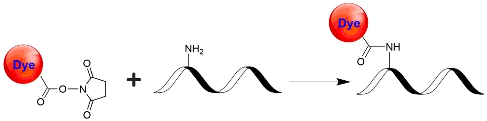
RuFluor™ succinimidyl ester
RuFluor™ succinimidyl ester is a highly water-soluble ruthenium complex that can be used in fluorescence polarization assays, time-resolved immunoassays, ECL immunoassays. Like other succinimidyl ester compounds, RuFluor™ succinimidyl ester label amines on biomolecules under mild conditions. The bioconjugates obtained from RuFluor™ succinimidyl ester are stable both chemically and photochemically. An electrochemiluminescence assays (ECL) is an antibody-based test designed to detect the presence of a biological target. In a typical ECL assay, the presence of an agent of interest creates a complex with two antibodies: one antibody is attached to a magnetic particle, the other antibody, in solution, is modified with a reporter molecule. The antigen/antibody complex is exposed to an electrode, which simultaneously attracts the magnetic bead and stimulates the detection molecule to emit light. The measurement of light is correlated with the presence of the specific antigen. ECL assays allow the detection of drugs and biomolecules in a wide range of molecular weights.
| Catalog | Size | Price | Quantity |
|---|---|---|---|
| 1520 | 1 mg | Price |
Physical properties
| Molecular weight | 1915.16 |
| Solvent | DMSO |
Usage and storage
| Intended use | Research Use Only (RUO) |
| Storage | Freeze (< -15 °C); Minimize light exposure |
Contact us
| Telephone | |
| Fax | |
| sales@aatbio.com | |
| International | See distributors |
| Bulk request | Inquire |
| Custom size | Inquire |
| Technical Support | Contact us |
| Request quotation | Request |
| Purchase order | Send to sales@aatbio.com |
| Shipping | Standard overnight for United States, inquire for international |
Page updated on July 16, 2026