logo
AAT Bioquest

Screen Quest™ Colorimetric Chloride Channel Assay Kit

NaI dose response was measured with Screen Quest™ Colorimetric Chloride Channel Assay Kit in a clear 96-well plate.
Ordering information
Price
Catalog Number
Unit Size
Quantity
Add to cart
Additional ordering information
Telephone1-800-990-8053
Fax1-800-609-2943
Emailsales@aatbio.com
InternationalSee distributors
Bulk requestInquire
Custom sizeInquire
ShippingStandard overnight for United States, inquire for international
Request quotation
Storage, safety and handling
H-phraseH303, H313, H333
Hazard symbolXN
Intended useResearch Use Only (RUO)
R-phraseR20, R21, R22
UNSPSC12352200
Related products
Screen Quest™ Membrane Potential Assay Kit *Orange Fluorescence*
Screen Quest™ Membrane Potential Assay Kit *Red Fluorescence*
Screen Quest™ 10X cell staining buffer with Phenol Red Plus™
Screen Quest™ 10X calcium assay buffer with Phenol Red Plus™
Screen Quest™ Luminometric Calcium Assay Kit
Screen Quest™ Fluo-8 Medium Removal Calcium Assay Kit *Optimized for Difficult Cell Lines*
Screen Quest™ Fluo-8 No Wash Calcium Assay Kit
Screen Quest™ Fura-2 No Wash Calcium Assay Kit
Screen Quest™ Rhod-4 No Wash Calcium Assay Kit *Medium Removal*
Screen Quest™ Rhod-4 No Wash Calcium Assay Kit
Screen Quest™ Fluorimetric MDR Assay Kit
Screen Quest™ Colorimetric ELISA cAMP Assay Kit
Screen Quest™ Fluorimetric ELISA cAMP Assay Kit
Screen Quest™ TR-FRET No Wash cAMP Assay Kit
Screen Quest™ Live Cell cAMP Assay Service Pack
Screen Quest™ Fluorimetric Fatty Acid Uptake Assay Kit
Screen Quest™ Fluorimetric Glucose Uptake Assay Kit
Screen Quest™ Colorimetric Glucose Uptake Assay Kit
Screen Quest™ HEK-CNGC-Amylin 3 Receptor Cells
Screen Quest™ HEK-CNGC-Cannabinoid Receptor 1 Cells
Screen Quest™ CHO-Ga16- Chemokine (C-C) Receptor 2B Cells
Screen Quest™ HEK-CNGC-Chemokine (C-X-C motif) Receptor 4 Cells
Screen Quest™ HEK-CNGC-Dopamine Receptor 1 (DRD1) Cells
Screen Quest™ HEK-CNGC-Glucagon-like Receptor 1 (GLP1R) Cells
Screen Quest™ HEK-CNGC-Opiate Receptor-like 1 (ORL1) Cells
Screen Quest™ Human Nociceptin Receptor Ga16 coupled CHO Cells (NOP-Ga16)
Screen Quest™ CHO-Gqi Chimera Cell line
Screen Quest™ CHO-Gqo Chimera Cell line
Screen Quest™ CHO-Gqz Chimera Cell line
Screen Quest™ CHO-Gqs Chimera Cell line
Screen Quest™ CHO-Ga16 Chimera Cell line
Screen Quest™ Fluo-4 No Wash Calcium Assay Kit
Screen Quest™ No Wash Potassium Channel Assay Kit
Screen Quest™ Calbryte-520 Probenecid-Free and Wash-Free Calcium Assay Kit
Screen Quest™ Calbryte-590 Probenecid-Free and Wash-Free Calcium Assay Kit
Show More (25)

OverviewpdfSDSpdfProtocol


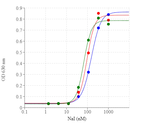
Chloride channels have a variety of important physiological and cellular functions that include regulation of pH, volume homeostasis, organic solute transport, cell migration, cell proliferation and differentiation. Chloride channels represent valuable drug targets. A number of chronic disease states such as cystic fibrosis and Bartter's syndrome are due to defects in chloride channel functions. However, the existing technologies for screening chloride channel modulators are a compromise between throughput, sensitivity and physiological relevance. Screen Quest™ Colorimetric Chloride Channel Assay Kit provides a sensitive and robust colorimetric method for studying chloride channels. The assay is based on our proprietary iodide indicator (Iodide Blue™) for measuring iodide concentration, as low as 30 nM of iodide was detected by this assay. Screen Quest™ Chloride Channel Assay Kit provides an optimized assay method for monitoring chloride channels. The assay can be performed in a convenient 96-well or 384-well microtiter-plate format and easily adapted to automation.

Platform


Absorbance microplate reader

Absorbance630, 380, or 405 nm
Recommended plateClear bottom

Components


Example protocol


AT A GLANCE

Protocol Summary
  1. Prepare cells
  2. Remove the growth medium
  3. Add I- Loading Buffer, treat cells with screening compounds
  4. Wash cells with DPBS buffer 3 times
  5. Lyse the cells with 1X lysis buffer
  6. Add equal volume of I-sensor (50 or 25 µL)
  7. Add 0.1X to 1X I- Sensor Enhancer (50 or 25 µL)
  8. Incubate at room temperature for 10 seconds to 10 minutes
  9. Monitor absorbance at 380 nm, 405 nm, or 630 nm 
Important      Warm all the reagents to room temperature before use.

CELL PREPARATION

For guidelines on cell sample preparation, please visit https://www.aatbio.com/resources/guides/cell-sample-preparation.html

PREPARATION OF STOCK SOLUTIONS

Unless otherwise noted, all unused stock solutions should be divided into single-use aliquots and stored at -20 °C after preparation. Avoid repeated freeze-thaw cycles.

Cell Lysis Buffer (1X)
Add the whole vial of 10X Cell Lysis Buffer (Component D) to 45 mL of sterile H2O and mix well.
Note     5 mL of 1X cell lysis buffer is enough for one plate. Store unused 1X cell lysis buffer at 4 °C.

PREPARATION OF WORKING SOLUTION

I- Sensor Enhancer solution (1X)
Add 50 µL of 100X Iodide sensor enhancer (Component B) to 5 mL of sterile H2O and mix well.
Note     1X I- Sensor Enhancer solution is not stable; use within 2 hours after the dilution. Note: Each cell line should be evaluated on an individual basis to determine the optimal dilution of I- Sensor Enhancer solution. We observed that 0.1X I- Sensor Enhancer solution works even better for some cell lines.

SAMPLE EXPERIMENTAL PROTOCOL

For iodide efflux assay
  1. Aspirate the growth medium from the cell plate.
  2. Add 100 µL/well (96-well plate) or 25 µL/well (384-well plate) of pre-warmed I- Loading Buffer (Component C) and incubate for 2 - 4 hours.
  3. Aspirate the Iodide Loading Buffer completely. Wash the cells with DPBS or HBSS at least 3 times.
  4. Treat the cells with agonist in HBSS buffer for 5 minutes.
    Note      For antagonists screen, incubate the compounds with I- loading buffer for at least an additional 30 min before the cells were washed with DPBS or HBSS buffer.
  5. Aspirate the supernatant.
  6. Lyse the cells by adding 50 µL/well (96-well plate) or 25 µL/well (384-well plate) of 1X cell lysis buffer.
  7. Perform the iodide assay. 

For iodide influx assay
  1. Aspirate the growth medium from the cell plate.
  2. Add 100 µL/well (96-well plate) or 25 µL/well (384-well plate) of pre-warmed I- Loading Buffer (Component C) with test compounds and incubate for 5 minutes.
  3. Aspirate the Iodide Loading Buffer completely. Wash the cells with HBSS 3 times.
  4. Lyse the cells by adding 50 µL/well (96-well plate) or 25 µL/well (384-well plate) of 1X cell lysis buffer.
  5. Perform the iodide assay. 

Run iodide assay
  1. Add 50 µL/well (96-well plate) or 25 µL/well (384-well plate) of Iodide Blue™ sensor (Component A) to the wells from your choice of iodide influx/efflux assay.
  2. Add 50 µL/well (96-well plate) or 25 µL/well (384-well plate) of 1X Iodide Sensor Enhancer solution into the mixture.
    Note     For some cell lines, you might need to dilute Enhancer solution down to 0.1X.
  3. Incubate at room temperature for 10 sec - 10 min.
    Note     Each cell line should be evaluated on an individual basis to determine the optimal incubation time.
    Note     The blue color may change to yellow within seconds to minutes due to the presence of a high concentration of iodide.
  4. Monitor absorbance at 630, 380, or 405 nm. 

Images


References


View all 44 references: Citation Explorer
Synthesis of 4-thiophen-2'-yl-1,4-dihydropyridines as potentiators of the CFTR chloride channel
Authors: Cateni F, Zacchigna M, Pedemonte N, Galietta LJ, Mazzei MT, Fossa P, Giampieri M, Mazzei M.
Journal: Bioorg Med Chem (2009): 7894
Genetically encoded optical sensors for monitoring of intracellular chloride and chloride-selective channel activity
Authors: Bregestovski P, Waseem T, Mukhtarov M.
Journal: Front Mol Neurosci (2009): 15
Chloride intracellular channel protein-4 functions in angiogenesis by supporting acidification of vacuoles along the intracellular tubulogenic pathway
Authors: Ulmasov B, Bruno J, Gordon N, Hartnett ME, Edwards JC.
Journal: Am J Pathol (2009): 1084
Statins and fenofibrate affect skeletal muscle chloride conductance in rats by differently impairing ClC-1 channel regulation and expression
Authors: Pierno S, Camerino GM, Cippone V, Roll and JF, Desaphy JF, De Luca A, Liantonio A, Bianco G, Kunic JD, George AL, Jr., Conte Camerino D.
Journal: Br J Pharmacol (2009): 1206
Spatiotemporal regulation of chloride intracellular channel protein CLIC4 by RhoA
Authors: Ponsioen B, van Zeijl L, Langeslag M, Berryman M, Littler D, Jalink K, Moolenaar WH.
Journal: Mol Biol Cell (2009): 4664
Chloride intracellular channel-4 is a determinant of native collateral formation in skeletal muscle and brain
Authors: Chalothorn D, Zhang H, Smith JE, Edwards JC, Faber JE.
Journal: Circ Res (2009): 89
Effects of Chinese herbal medicine combined with He-Ne laser on lipoperoxide and superoxide dismutase in chloasma patients
Authors: Wu YH, Li QL, Yang XW.
Journal: J Tradit Chin Med (2009): 163
Chloride intracellular channel 1 regulates osteoblast differentiation
Authors: Yang JY, Jung JY, Cho SW, Choi HJ, Kim SW, Kim SY, Kim HJ, Jang CH, Lee MG, Han J, Shin CS.
Journal: Bone (2009): 1175
Two novel CLCN2 mutations accelerating chloride channel deactivation are associated with idiopathic generalized epilepsy
Authors: Saint-Martin C, Gauvain G, Teodorescu G, Gourfinkel-An I, Fedirko E, Weber YG, Maljevic S, Ernst JP, Garcia-Olivares J, Fahlke C, Nabbout R, LeGuern E, Lerche H, Poncer JC, Depienne C.
Journal: Hum Mutat (2009): 397
Identification of metastasis-associated proteins involved in gallbladder carcinoma metastasis by proteomic analysis and functional exploration of chloride intracellular channel 1
Authors: Wang JW, Peng SY, Li JT, Wang Y, Zhang ZP, Cheng Y, Cheng DQ, Weng WH, Wu XS, Fei XZ, Quan ZW, Li JY, Li SG, Liu YB.
Journal: Cancer Lett (2009): 71